• 文献检索
  • 文档翻译
  • 深度研究
  • 学术资讯
  • Suppr Zotero 插件Zotero 插件
  • 邀请有礼
  • 套餐&价格
  • 历史记录
应用&插件
Suppr Zotero 插件Zotero 插件浏览器插件Mac 客户端Windows 客户端微信小程序
定价
高级版会员购买积分包购买API积分包
服务
文献检索文档翻译深度研究API 文档MCP 服务
关于我们
关于 Suppr公司介绍联系我们用户协议隐私条款
关注我们

Suppr 超能文献

核心技术专利:CN118964589B侵权必究
粤ICP备2023148730 号-1Suppr @ 2026

文献检索

告别复杂PubMed语法,用中文像聊天一样搜索,搜遍4000万医学文献。AI智能推荐,让科研检索更轻松。

立即免费搜索

文件翻译

保留排版,准确专业,支持PDF/Word/PPT等文件格式,支持 12+语言互译。

免费翻译文档

深度研究

AI帮你快速写综述,25分钟生成高质量综述,智能提取关键信息,辅助科研写作。

立即免费体验

高场 MRI 在猪的骨软骨病易发病部位显示软骨管的组织学证实和生物学意义。

Histological confirmation and biological significance of cartilage canals demonstrated using high field MRI in swine at predilection sites of osteochondrosis.

机构信息

Department of Veterinary Population Medicine, College of Veterinary Medicine, University of Minnesota, St. Paul, Minnesota.

出版信息

J Orthop Res. 2013 Dec;31(12):2006-12. doi: 10.1002/jor.22449. Epub 2013 Aug 12.

DOI:10.1002/jor.22449
PMID:23939946
原文链接:https://pmc.ncbi.nlm.nih.gov/articles/PMC4034470/
Abstract

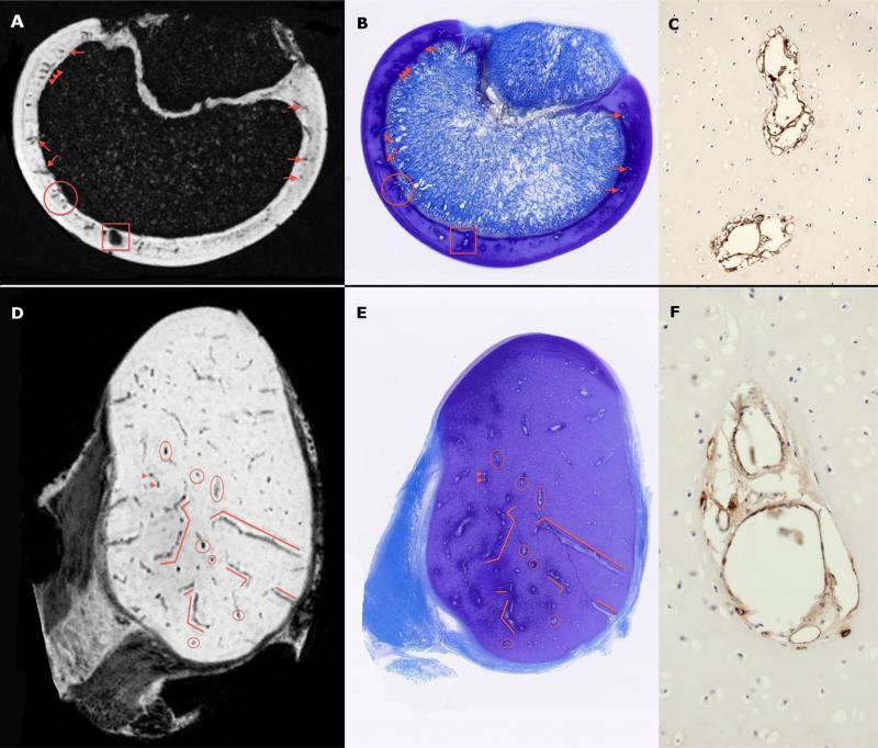
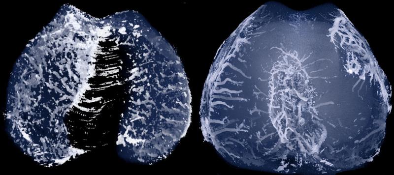
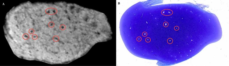
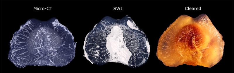
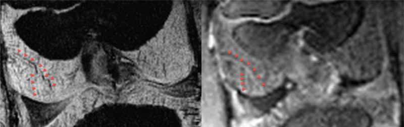
Cartilage canal vessels in epiphyseal cartilage have a pivotal role in the pathogenesis of osteochondrosis/osteochondritis dissecans. The present study aimed to validate high field magnetic resonance imaging (MRI) methods to visualize these vessels in young pigs. Osteochondral samples from the distal femur and distal humerus (predilection sites of osteochondrosis) of piglets were imaged post-mortem: (1) using susceptibility-weighted imaging (SWI) in an MRI scanner, followed by histological evaluation; and (2) after barium perfusion using µCT, followed by clearing techniques. In addition, both stifle joints of a 25-day-old piglet were imaged in vivo using SWI and gadolinium enhanced T1-weighted MRI, after which distal femoral samples were harvested and evaluated using µCT and histology. Histological sections were compared to corresponding MRI slices, and three-dimensional visualizations of vessels identified using MRI were compared to those obtained using µCT and to the cleared specimens. Vessels contained in cartilage canals were identified using MRI, both ex vivo and in vivo; their locations matched those observed in the histological sections, µCT images, and cleared specimens of barium-perfused tissues. The ability to visualize cartilage canal blood vessels by MRI, without using a contrast agent, will allow future longitudinal studies to evaluate their role in developmental orthopedic disease.

摘要

骺软骨内的软骨管血管在骨软骨病/剥脱性骨软骨炎的发病机制中起着关键作用。本研究旨在验证高磁场磁共振成像(MRI)方法在年轻猪中可视化这些血管的能力。死后对仔猪的股骨远端和肱骨远端(骨软骨病的易发病部位)的骨软骨样本进行成像:(1)在 MRI 扫描仪中使用磁敏感加权成像(SWI),然后进行组织学评估;(2)在使用微 CT 进行钡灌注后,然后使用透明技术。此外,在使用 SWI 和钆增强 T1 加权 MRI 对一只 25 天大的仔猪的两个膝关节进行了活体成像,之后采集并使用微 CT 和组织学评估了股骨远端样本。将组织学切片与相应的 MRI 切片进行比较,将 MRI 识别的血管的三维可视化与微 CT 获得的和透明样本进行比较。在体外用和体内用 MRI 都可以识别软骨管内的血管;它们的位置与在组织学切片、微 CT 图像和钡灌注组织的透明标本中观察到的位置相匹配。无需造影剂即可通过 MRI 可视化软骨管血管的能力,将允许未来的纵向研究评估它们在发育性骨科疾病中的作用。

https://cdn.ncbi.nlm.nih.gov/pmc/blobs/a734/4034470/6f433e4fd45f/nihms-572241-f0005.jpg
https://cdn.ncbi.nlm.nih.gov/pmc/blobs/a734/4034470/aa9754305188/nihms-572241-f0001.jpg
https://cdn.ncbi.nlm.nih.gov/pmc/blobs/a734/4034470/7b0d6dc40250/nihms-572241-f0002.jpg
https://cdn.ncbi.nlm.nih.gov/pmc/blobs/a734/4034470/8216b46fd2dd/nihms-572241-f0003.jpg
https://cdn.ncbi.nlm.nih.gov/pmc/blobs/a734/4034470/d4cda5756171/nihms-572241-f0004.jpg
https://cdn.ncbi.nlm.nih.gov/pmc/blobs/a734/4034470/6f433e4fd45f/nihms-572241-f0005.jpg

相似文献

1
Histological confirmation and biological significance of cartilage canals demonstrated using high field MRI in swine at predilection sites of osteochondrosis.高场 MRI 在猪的骨软骨病易发病部位显示软骨管的组织学证实和生物学意义。
J Orthop Res. 2013 Dec;31(12):2006-12. doi: 10.1002/jor.22449. Epub 2013 Aug 12.
2
Novel Application of Magnetic Resonance Imaging Demonstrates Characteristic Differences in Vasculature at Predilection Sites of Osteochondritis Dissecans.磁共振成像的新应用显示了剥脱性骨软骨炎好发部位血管系统的特征差异。
Am J Sports Med. 2015 Oct;43(10):2522-7. doi: 10.1177/0363546515596410. Epub 2015 Aug 18.
3
Susceptibility weighted imaging of cartilage canals in porcine epiphyseal growth cartilage ex vivo and in vivo.猪骨骺生长软骨中软骨管的体外和体内磁共振敏感性加权成像
Magn Reson Med. 2014 Jun;71(6):2197-205. doi: 10.1002/mrm.24863. Epub 2013 Jul 15.
4
Validation of the ultrasonographic assessment of the femoral trochlea epiphyseal cartilage in foals at osteochondrosis predilected sites with magnetic resonance imaging and histology.通过磁共振成像和组织学对马驹骨软骨病好发部位股骨滑车骨骺软骨超声评估的验证。
Equine Vet J. 2017 Nov;49(6):821-828. doi: 10.1111/evj.12698. Epub 2017 Jun 19.
5
Differences in the vascular tree of the femoral trochlear growth cartilage at osteochondrosis-susceptible sites in foals revealed by SWI 3T MRI.3T磁共振弹性成像(SWI)显示马驹易患骨软骨病部位股骨滑车生长软骨血管树的差异。
J Orthop Res. 2016 Sep;34(9):1539-46. doi: 10.1002/jor.23149. Epub 2016 Jan 21.
6
Surgical induction, histological evaluation, and MRI identification of cartilage necrosis in the distal femur in goats to model early lesions of osteochondrosis.通过手术诱导、组织学评估和MRI识别山羊股骨远端软骨坏死,以模拟骨软骨病的早期病变。
Osteoarthritis Cartilage. 2015 Feb;23(2):300-7. doi: 10.1016/j.joca.2014.11.009. Epub 2014 Nov 15.
7
Cartilage canals and lesions typical of osteochondrosis in growth cartilages from the distal part of the humerus of newborn pigs.
Vet Rec. 1991 Mar 9;128(10):221-8. doi: 10.1136/vr.128.10.221.
8
Transection of vessels in epiphyseal cartilage canals leads to osteochondrosis and osteochondrosis dissecans in the femoro-patellar joint of foals; a potential model of juvenile osteochondritis dissecans.骺软骨管中的血管横断可导致驹膝关节的骨软骨病和骨软骨骨软骨病;青少年剥脱性骨软骨炎的潜在模型。
Osteoarthritis Cartilage. 2013 May;21(5):730-8. doi: 10.1016/j.joca.2013.02.005. Epub 2013 Feb 18.
9
Ischemic necrosis of cartilage in spontaneous and experimental lesions of osteochondrosis.骨软骨病自发性及实验性病变中软骨的缺血性坏死
J Orthop Res. 1991 May;9(3):317-29. doi: 10.1002/jor.1100090303.
10
Epiphyseal cartilage canal blood supply to the tarsus of foals and relationship to osteochondrosis.马驹跗骨的骨骺软骨管血液供应及其与骨软骨病的关系
Equine Vet J. 2008 Jan;40(1):30-9. doi: 10.2746/042516407X239836.

引用本文的文献

1
Magnetic resonance-enhanced high-resolution three-dimensional water-selective cartilage sequence visualization of hip vessels in children.磁共振增强高分辨率三维水选择性软骨序列在儿童髋关节血管中的可视化。
Int Orthop. 2024 Jun;48(6):1391-1399. doi: 10.1007/s00264-023-05883-x. Epub 2023 Jul 14.
2
Ultra-high-field MRI of postmortem human fetal wrist joints: initial experience.超高场磁共振成像在后胎儿人类腕关节:初步经验。
Eur Radiol Exp. 2023 Jun 5;7(1):28. doi: 10.1186/s41747-023-00341-0.
3
A method for labelling lesions for machine learning and some new observations on osteochondrosis in computed tomographic scans of four pig joints.

本文引用的文献

1
Dynamically applied B1+ shimming solutions for non-contrast enhanced renal angiography at 7.0 Tesla.7.0 特斯拉下非对比增强肾血管造影的动态 B1+匀场解决方案。
Magn Reson Med. 2013 Jan;69(1):114-26. doi: 10.1002/mrm.24237. Epub 2012 Mar 22.
2
Simultaneous bilateral hip joint imaging at 7 Tesla using fast transmit B₁ shimming methods and multichannel transmission - a feasibility study.在 7 特斯拉场强下使用快速发射 B₁匀场方法和多通道传输进行双侧髋关节同步成像-可行性研究。
NMR Biomed. 2012 Oct;25(10):1202-8. doi: 10.1002/nbm.2779. Epub 2012 Feb 7.
3
Susceptibility-weighted imaging: technical aspects and clinical applications, part 1.
一种用于机器学习的病变标记方法及对四只猪关节 CT 扫描中骨软骨病的一些新观察。
BMC Vet Res. 2022 Aug 31;18(1):328. doi: 10.1186/s12917-022-03426-x.
4
Vascularity of the early post-natal human distal femoral chondroepiphysis: Quantitative MRI analysis.出生后早期人类股骨远端软骨骨骺的血管分布:定量MRI分析
J Child Orthop. 2022 Apr;16(2):152-158. doi: 10.1177/18632521221084179. Epub 2022 Apr 30.
5
Epiphyseal cartilage canal architecture and extracellular matrix remodeling in mucopolysaccharidosis VII dogs at the onset of postnatal growth.黏多糖贮积症 VII 型犬在出生后生长开始时的骺软骨管结构和细胞外基质重塑。
Connect Tissue Res. 2021 Nov;62(6):698-708. doi: 10.1080/03008207.2020.1865939. Epub 2021 Jan 11.
6
Three-Dimensional Quantitative Magnetic Resonance Imaging of Epiphyseal Cartilage Vascularity Using Vessel Image Features: New Insights into Juvenile Osteochondritis Dissecans.利用血管图像特征对骨骺软骨血管进行三维定量磁共振成像:对青少年剥脱性骨软骨炎的新见解
JB JS Open Access. 2019 Dec 5;4(4). doi: 10.2106/JBJS.OA.19.00031. eCollection 2019 Oct-Dec.
7
Quantitative susceptibility mapping detects neovascularization of the epiphyseal cartilage after ischemic injury in a piglet model of legg-calvé-perthes disease.定量磁化率映射检测 legg-calvé-perthes 病猪模型缺血性损伤后骺软骨新生血管形成。
J Magn Reson Imaging. 2019 Jul;50(1):106-113. doi: 10.1002/jmri.26552. Epub 2018 Dec 17.
8
Vessel architecture in human knee cartilage in children: an in vivo susceptibility-weighted imaging study at 7 T.人体膝关节软骨中的血管结构:7T 下体内磁共振敏感加权成像研究。
Eur Radiol. 2018 Aug;28(8):3384-3392. doi: 10.1007/s00330-017-5290-1. Epub 2018 Feb 26.
9
Non-linear optical microscopy of cartilage canals in the distal femur of young pigs may reveal the cause of articular osteochondrosis.对幼猪股骨远端软骨管进行非线性光学显微镜检查可能揭示关节骨软骨病的病因。
BMC Vet Res. 2017 Aug 22;13(1):270. doi: 10.1186/s12917-017-1197-y.
10
Quantitative susceptibility mapping detects abnormalities in cartilage canals in a goat model of preclinical osteochondritis dissecans.定量磁化率成像在临床前剥脱性骨软骨炎山羊模型中检测软骨管异常。
Magn Reson Med. 2017 Mar;77(3):1276-1283. doi: 10.1002/mrm.26214. Epub 2016 Mar 28.
磁敏感加权成像:技术要点与临床应用,第1部分
AJNR Am J Neuroradiol. 2009 Jan;30(1):19-30. doi: 10.3174/ajnr.A1400. Epub 2008 Nov 27.
4
Epiphyseal cartilage canal blood supply to the distal femur of foals.马驹股骨远端的骨骺软骨管血液供应。
Equine Vet J. 2008 Jul;40(5):433-9. doi: 10.2746/042516408X300269.
5
Epiphyseal cartilage canal blood supply to the tarsus of foals and relationship to osteochondrosis.马驹跗骨的骨骺软骨管血液供应及其与骨软骨病的关系
Equine Vet J. 2008 Jan;40(1):30-9. doi: 10.2746/042516407X239836.
6
Internal fixation of juvenile osteochondritis dissecans lesions of the knee.青少年膝关节剥脱性骨软骨炎病变的内固定术
Am J Sports Med. 2007 May;35(5):712-8. doi: 10.1177/0363546506296608. Epub 2007 Mar 2.
7
Gadolinium-based MR contrast agents and nephrogenic systemic fibrosis.钆基磁共振造影剂与肾源性系统性纤维化
Radiology. 2007 Mar;242(3):647-9. doi: 10.1148/radiol.2423061640. Epub 2007 Jan 9.
8
Cartilage Canals.软骨管道
J Anat. 1933 Oct;68(Pt 1):45-64.
9
B(1) destructive interferences and spatial phase patterns at 7 T with a head transceiver array coil.使用头部收发器阵列线圈在7T时的B(1) 相消干涉和空间相位模式。
Magn Reson Med. 2005 Dec;54(6):1503-18. doi: 10.1002/mrm.20708.
10
The role of cartilage canals in endochondral and perichondral bone formation: are there similarities between these two processes?软骨管在软骨内成骨和软骨膜下骨形成中的作用:这两个过程之间存在相似之处吗?
J Anat. 2005 Apr;206(4):359-72. doi: 10.1111/j.1469-7580.2005.00404.x.