• 文献检索
  • 文档翻译
  • 深度研究
  • 学术资讯
  • Suppr Zotero 插件Zotero 插件
  • 邀请有礼
  • 套餐&价格
  • 历史记录
应用&插件
Suppr Zotero 插件Zotero 插件浏览器插件Mac 客户端Windows 客户端微信小程序
定价
高级版会员购买积分包购买API积分包
服务
文献检索文档翻译深度研究API 文档MCP 服务
关于我们
关于 Suppr公司介绍联系我们用户协议隐私条款
关注我们

Suppr 超能文献

核心技术专利:CN118964589B侵权必究
粤ICP备2023148730 号-1Suppr @ 2026

文献检索

告别复杂PubMed语法,用中文像聊天一样搜索,搜遍4000万医学文献。AI智能推荐,让科研检索更轻松。

立即免费搜索

文件翻译

保留排版,准确专业,支持PDF/Word/PPT等文件格式,支持 12+语言互译。

免费翻译文档

深度研究

AI帮你快速写综述,25分钟生成高质量综述,智能提取关键信息,辅助科研写作。

立即免费体验

氧化铜纳米颗粒诱导人肝癌细胞线粒体介导的细胞凋亡。

Copper oxide nanoparticles induced mitochondria mediated apoptosis in human hepatocarcinoma cells.

机构信息

Department of Zoology, College of Science, King Saud University, Riyadh, Saudi Arabia.

出版信息

PLoS One. 2013 Aug 5;8(8):e69534. doi: 10.1371/journal.pone.0069534. Print 2013.

DOI:10.1371/journal.pone.0069534
PMID:23940521
原文链接:https://pmc.ncbi.nlm.nih.gov/articles/PMC3734287/
Abstract

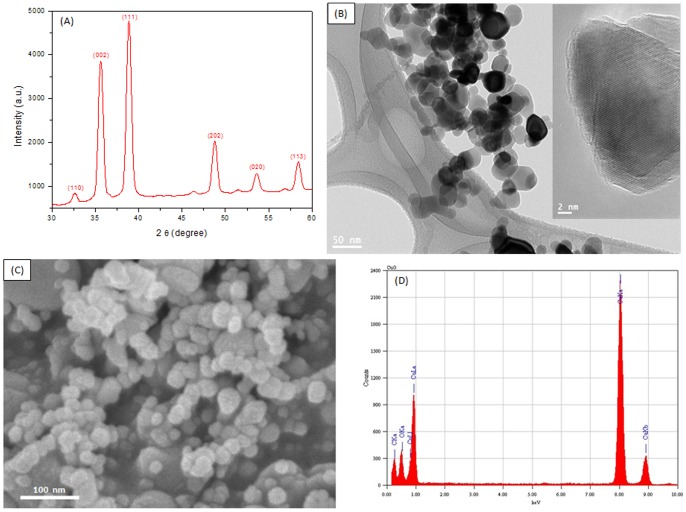
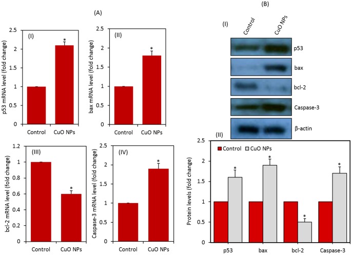
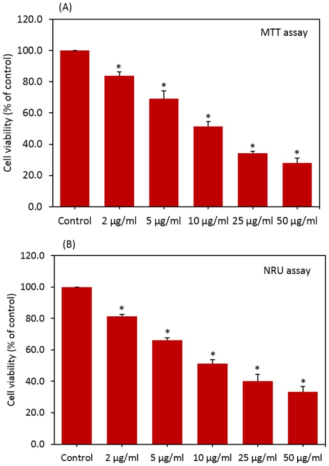
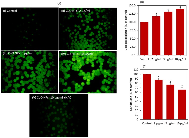
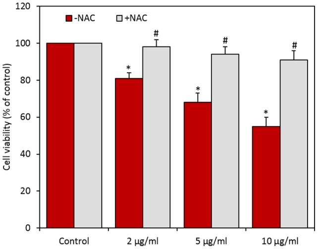
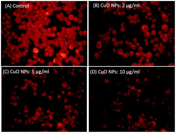
Copper oxide nanoparticles (CuO NPs) are heavily utilized in semiconductor devices, gas sensor, batteries, solar energy converter, microelectronics and heat transfer fluids. It has been reported that liver is one of the target organs for nanoparticles after they gain entry into the body through any of the possible routes. Recent studies have shown cytotoxic response of CuO NPs in liver cells. However, the underlying mechanism of apoptosis in liver cells due to CuO NPs exposure is largely lacking. We explored the possible mechanisms of apoptosis induced by CuO NPs in human hepatocellular carcinoma HepG2 cells. Prepared CuO NPs were spherical in shape with a smooth surface and had an average diameter of 22 nm. CuO NPs (concentration range 2-50 µg/ml) were found to induce cytotoxicity in HepG2 cells in dose-dependent manner, which was likely to be mediated through reactive oxygen species generation and oxidative stress. Tumor suppressor gene p53 and apoptotic gene caspase-3 were up-regulated due to CuO NPs exposure. Decrease in mitochondrial membrane potential with a concomitant increase in the gene expression of bax/bcl2 ratio suggested that mitochondria mediated pathway involved in CuO NPs induced apoptosis. This study has provided valuable insights into the possible mechanism of apoptosis caused by CuO NPs at in vitro level. Underlying mechanism(s) of apoptosis due to CuO NPs exposure should be further invested at in vivo level.

摘要

氧化铜纳米粒子(CuO NPs)在半导体器件、气体传感器、电池、太阳能转换器、微电子和热传递流体中得到了广泛的应用。据报道,纳米粒子通过任何可能的途径进入体内后,肝脏是其靶向器官之一。最近的研究表明,CuO NPs 在肝细胞中具有细胞毒性反应。然而,由于 CuO NPs 暴露导致肝细胞凋亡的潜在机制在很大程度上尚不清楚。我们探索了 CuO NPs 诱导人肝癌 HepG2 细胞凋亡的可能机制。制备的 CuO NPs 呈球形,表面光滑,平均直径为 22nm。研究发现,CuO NPs(浓度范围为 2-50μg/ml)以剂量依赖的方式诱导 HepG2 细胞毒性,这可能是通过活性氧生成和氧化应激介导的。肿瘤抑制基因 p53 和凋亡基因 caspase-3 因 CuO NPs 的暴露而上调。线粒体膜电位的下降伴随着 bax/bcl2 比值的基因表达增加,提示线粒体介导的途径参与了 CuO NPs 诱导的细胞凋亡。本研究为 CuO NPs 在体外引起细胞凋亡的可能机制提供了有价值的见解。应在体内水平进一步研究 CuO NPs 暴露引起细胞凋亡的潜在机制。

https://cdn.ncbi.nlm.nih.gov/pmc/blobs/fb01/3734287/8cf9831bc3c9/pone.0069534.g006.jpg
https://cdn.ncbi.nlm.nih.gov/pmc/blobs/fb01/3734287/0ed295ab5cd1/pone.0069534.g001.jpg
https://cdn.ncbi.nlm.nih.gov/pmc/blobs/fb01/3734287/3d20a128296d/pone.0069534.g002.jpg
https://cdn.ncbi.nlm.nih.gov/pmc/blobs/fb01/3734287/c2dc96adc801/pone.0069534.g003.jpg
https://cdn.ncbi.nlm.nih.gov/pmc/blobs/fb01/3734287/2357cb9f6dc9/pone.0069534.g004.jpg
https://cdn.ncbi.nlm.nih.gov/pmc/blobs/fb01/3734287/161d393b28c1/pone.0069534.g005.jpg
https://cdn.ncbi.nlm.nih.gov/pmc/blobs/fb01/3734287/8cf9831bc3c9/pone.0069534.g006.jpg

相似文献

1
Copper oxide nanoparticles induced mitochondria mediated apoptosis in human hepatocarcinoma cells.氧化铜纳米颗粒诱导人肝癌细胞线粒体介导的细胞凋亡。
PLoS One. 2013 Aug 5;8(8):e69534. doi: 10.1371/journal.pone.0069534. Print 2013.
2
Enhanced reactive oxygen species overexpression by CuO nanoparticles in poorly differentiated hepatocellular carcinoma cells.氧化铜纳米颗粒增强低分化肝癌细胞内活性氧的过度表达。
Nanoscale. 2015 Feb 7;7(5):1820-9. doi: 10.1039/c4nr05843g.
3
Copper ferrite nanoparticle-induced cytotoxicity and oxidative stress in human breast cancer MCF-7 cells.铜铁氧体纳米颗粒诱导人乳腺癌 MCF-7 细胞的细胞毒性和氧化应激。
Colloids Surf B Biointerfaces. 2016 Jun 1;142:46-54. doi: 10.1016/j.colsurfb.2016.02.043. Epub 2016 Feb 22.
4
Amphipathic silica nanoparticles induce cytotoxicity through oxidative stress mediated and p53 dependent apoptosis pathway in human liver cell line HL-7702 and rat liver cell line BRL-3A.两亲性二氧化硅纳米颗粒通过氧化应激介导的和p53依赖性凋亡途径在人肝细胞系HL-7702和大鼠肝细胞系BRL-3A中诱导细胞毒性。
Colloids Surf B Biointerfaces. 2016 Sep 1;145:232-240. doi: 10.1016/j.colsurfb.2016.05.006. Epub 2016 May 5.
5
Pomegranate Juice Diminishes The Mitochondria-Dependent Cell Death And NF-kB Signaling Pathway Induced By Copper Oxide Nanoparticles On Liver And Kidneys Of Rats.石榴汁可减轻氧化铜纳米颗粒诱导的大鼠肝、肾细胞中线粒体依赖的细胞死亡和 NF-kB 信号通路。
Int J Nanomedicine. 2019 Nov 15;14:8905-8922. doi: 10.2147/IJN.S229461. eCollection 2019.
6
Can CuO nanoparticles lead to epigenetic regulation of antioxidant enzyme system?氧化铜纳米颗粒会导致抗氧化酶系统的表观遗传调控吗?
J Appl Toxicol. 2017 Jan;37(1):84-91. doi: 10.1002/jat.3392. Epub 2016 Sep 30.
7
CuO nanoparticles induce cytotoxicity and apoptosis in human K562 cancer cell line via mitochondrial pathway, through reactive oxygen species and P53.氧化铜纳米颗粒通过线粒体途径,经由活性氧和P53,在人K562癌细胞系中诱导细胞毒性和凋亡。
Iran J Basic Med Sci. 2015 Oct;18(10):993-1000.
8
Oxidative stress contributes to cobalt oxide nanoparticles-induced cytotoxicity and DNA damage in human hepatocarcinoma cells.氧化应激导致氧化钴纳米颗粒诱导人肝癌细胞的细胞毒性和 DNA 损伤。
Int J Nanomedicine. 2013;8:189-199. doi: 10.2147/IJN.S37924. Epub 2013 Jan 8.
9
Nickel oxide nanoparticles exert cytotoxicity via oxidative stress and induce apoptotic response in human liver cells (HepG2).氧化镍纳米颗粒通过氧化应激对人肝细胞(HepG2)产生细胞毒性,并诱导其发生凋亡反应。
Chemosphere. 2013 Nov;93(10):2514-22. doi: 10.1016/j.chemosphere.2013.09.047. Epub 2013 Oct 15.
10
Influence of silica nanoparticles on cadmium-induced cytotoxicity, oxidative stress, and apoptosis in human liver HepG2 cells.二氧化硅纳米颗粒对镉诱导的人肝癌 HepG2 细胞毒性、氧化应激和细胞凋亡的影响。
Environ Toxicol. 2020 May;35(5):599-608. doi: 10.1002/tox.22895. Epub 2020 Jan 6.

引用本文的文献

1
Epothilone B from Aspergillus fumigatus with a strong antiproliferative and anti-tubulin polymerizing activities; apoptosis, and cell cycle analyses.来自烟曲霉的埃坡霉素B具有强大的抗增殖和抗微管蛋白聚合活性;凋亡及细胞周期分析。
BMC Microbiol. 2025 Jun 26;25(1):367. doi: 10.1186/s12866-025-04086-1.
2
Influence of capping agents on physicochemical properties and leukemic cytotoxicity of copper oxide nanoparticles biosynthesized using Caesalpinia sappan extract.封端剂对使用苏木提取物生物合成的氧化铜纳米颗粒的物理化学性质和白血病细胞毒性的影响。
PLoS One. 2025 Jun 26;20(6):e0326791. doi: 10.1371/journal.pone.0326791. eCollection 2025.
3

本文引用的文献

1
Genotoxic effects of copper oxide nanoparticles in Neuro 2A cell cultures.氧化铜纳米颗粒对 Neuro 2A 细胞培养的遗传毒性作用。
Sci Total Environ. 2012 Dec 15;441:117-24. doi: 10.1016/j.scitotenv.2012.09.065. Epub 2012 Nov 6.
2
Copper(II) oxide nanoparticles penetrate into HepG2 cells, exert cytotoxicity via oxidative stress and induce pro-inflammatory response.氧化铜纳米颗粒穿透 HepG2 细胞,通过氧化应激发挥细胞毒性作用,并诱导促炎反应。
Nanoscale. 2012 Nov 21;4(22):7168-84. doi: 10.1039/c2nr31785k.
3
Nanosized copper oxide induces apoptosis through oxidative stress in podocytes.
Research Trends on Nanomaterials and Hepatocellular Carcinoma From 1999 to 2024: A Bibliometric Analysis.
1999年至2024年纳米材料与肝细胞癌的研究趋势:文献计量分析
Drug Des Devel Ther. 2025 May 16;19:3949-3970. doi: 10.2147/DDDT.S516647. eCollection 2025.
4
Cuproplasia and cuproptosis in hepatocellular carcinoma: mechanisms, relationship and potential role in tumor microenvironment and treatment.肝细胞癌中的铜代谢异常与铜死亡:机制、关系及其在肿瘤微环境和治疗中的潜在作用
Cancer Cell Int. 2025 Apr 9;25(1):137. doi: 10.1186/s12935-025-03683-4.
5
The Composition of the Dispersion Medium Determines the Antibacterial Properties of Copper (II) Oxide Nanoparticles Against Bacteria.分散介质的组成决定了氧化铜(II)纳米颗粒对细菌的抗菌性能。
Nanomaterials (Basel). 2025 Mar 20;15(6):469. doi: 10.3390/nano15060469.
6
Ultrasonic-assisted europium decorated cuprous oxide nanoparticles: exploring their photothermal capabilities and antioxidant properties for biomedical applications.超声辅助铕修饰的氧化亚铜纳米颗粒:探索其在生物医学应用中的光热性能和抗氧化性能
RSC Adv. 2025 Mar 4;15(9):6984-6993. doi: 10.1039/d4ra08914f. eCollection 2025 Feb 26.
7
Differential Cytotoxicity and Inflammatory Responses to Particulate Matter Components in Airway Structural Cells.气道结构细胞对颗粒物成分的细胞毒性差异及炎症反应
Int J Mol Sci. 2025 Jan 20;26(2):830. doi: 10.3390/ijms26020830.
8
The potential of copper oxide nanoparticles in nanomedicine: A comprehensive review.氧化铜纳米颗粒在纳米医学中的潜力:综述
Biotechnol Notes. 2024 Jun 5;5:80-99. doi: 10.1016/j.biotno.2024.06.001. eCollection 2024.
9
Navigating the microalgal maze: a comprehensive review of recent advances and future perspectives in biological networks.探索微藻迷宫:生物网络最新进展与未来展望的全面综述。
Planta. 2024 Oct 5;260(5):114. doi: 10.1007/s00425-024-04543-7.
10
Comparative study of influence of Cu, CuO nanoparticles and Cu on rainbow trout (Oncorhynchus mykiss W.) spermatozoa.铜、氧化铜纳米粒子和铜对虹鳟(Oncorhynchus mykiss W.)精子影响的比较研究。
Sci Rep. 2024 Sep 27;14(1):22242. doi: 10.1038/s41598-024-72956-1.
纳米氧化铜通过氧化应激诱导足细胞凋亡。
Arch Toxicol. 2013 Jun;87(6):1067-73. doi: 10.1007/s00204-012-0925-0. Epub 2012 Aug 18.
4
Copper oxide nanoparticles can induce toxicity to the freshwater shredder Allogamus ligonifer.氧化铜纳米颗粒会对淡水水丝蚓造成毒性。
Chemosphere. 2012 Nov;89(9):1142-50. doi: 10.1016/j.chemosphere.2012.06.001. Epub 2012 Jun 30.
5
Tumor suppression in the absence of p53-mediated cell-cycle arrest, apoptosis, and senescence.在没有 p53 介导的细胞周期阻滞、细胞凋亡和衰老的情况下抑制肿瘤。
Cell. 2012 Jun 8;149(6):1269-83. doi: 10.1016/j.cell.2012.04.026.
6
Accumulation and toxicity of copper oxide nanoparticles in the digestive gland of Mytilus galloprovincialis.铜氧化纳米颗粒在贻贝消化腺中的积累和毒性。
Aquat Toxicol. 2012 Aug 15;118-119:72-79. doi: 10.1016/j.aquatox.2012.03.017. Epub 2012 Apr 3.
7
Zinc oxide nanoparticles induce oxidative DNA damage and ROS-triggered mitochondria mediated apoptosis in human liver cells (HepG2).氧化锌纳米颗粒诱导人肝细胞(HepG2)氧化 DNA 损伤和 ROS 触发的线粒体介导的细胞凋亡。
Apoptosis. 2012 Aug;17(8):852-70. doi: 10.1007/s10495-012-0705-6.
8
Zinc oxide nanoparticles selectively induce apoptosis in human cancer cells through reactive oxygen species.氧化锌纳米颗粒通过活性氧选择性诱导人癌细胞凋亡。
Int J Nanomedicine. 2012;7:845-57. doi: 10.2147/IJN.S29129. Epub 2012 Feb 21.
9
Nickel oxide nanoparticles induce cytotoxicity, oxidative stress and apoptosis in cultured human cells that is abrogated by the dietary antioxidant curcumin.氧化镍纳米颗粒诱导培养的人细胞产生细胞毒性、氧化应激和细胞凋亡,膳食抗氧化剂姜黄素可阻断此作用。
Food Chem Toxicol. 2012 Mar;50(3-4):641-7. doi: 10.1016/j.fct.2012.01.017. Epub 2012 Jan 18.
10
Apoptosis induction by silica nanoparticles mediated through reactive oxygen species in human liver cell line HepG2.矽纳米颗粒通过活性氧物种诱导人肝癌细胞株 HepG2 细胞凋亡。
Toxicol Appl Pharmacol. 2012 Mar 1;259(2):160-8. doi: 10.1016/j.taap.2011.12.020. Epub 2012 Jan 8.