Department of Laboratory Animal Facility, CSIR-Indian Institute of Chemical Biology, Kolkata, India.
PLoS One. 2013 Aug 5;8(8):e70528. doi: 10.1371/journal.pone.0070528. Print 2013.
To evaluate the effects of pirfenidone nanoparticles on corneal re-epithelialization and scarring, major clinical challenges after alkali burn.
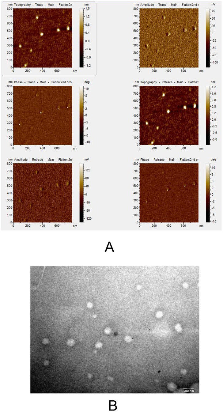
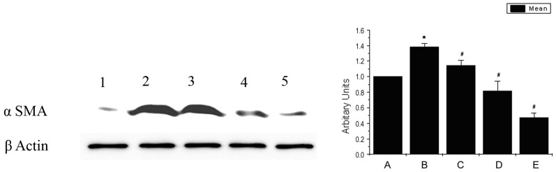
Effect of pirfenidone on collagen I and α-smooth muscle actin (α-SMA) synthesis by TGFβ induced primary corneal fibroblast cells was evaluated by immunoblotting and immunocytochemistry. Pirfenidone loaded poly (lactide-co-glycolide) (PLGA) nanoparticles were prepared, characterized and their cellular entry was examined in primary corneal fibroblast cells by fluorescence microscopy. Alkali burn was induced in one eye of Sprague Dawley rats followed by daily topical treatment with free pirfenidone, pirfenidone nanoparticles or vehicle. Corneal re-epithelialization was assessed daily by flourescein dye test; absence of stained area indicated complete re-epithelialization and the time for complete re-epithelialization was determined. Corneal haze was assessed daily for 7 days under slit lamp microscope and graded using a standard method. After 7 days, collagen I deposition in the superficial layer of cornea was examined by immunohistochemistry.
Pirfenidone prevented (P<0.05) increase in TGF β induced collagen I and α-SMA synthesis by corneal fibroblasts in a dose dependent manner. Pirfenidone could be loaded successfully within PLGA nanoparticles, which entered the corneal fibroblasts within 5 minutes. Pirfenidone nanoparticles but not free pirfenidone significantly (P<0.05) reduced collagen I level, corneal haze and the time for corneal re-epithelialization following alkali burn.
Pirfenidone decreases collagen synthesis and prevents myofibroblast formation. Pirfenidone nanoparticles improve corneal wound healing and prevent fibrosis. Pirfenidone nanoparticles are of potential value in treating corneal chemical burns and other corneal fibrotic diseases.
评估吡非尼酮纳米粒对角膜再上皮化和瘢痕形成的影响,这是碱烧伤后主要的临床挑战。
通过免疫印迹和免疫细胞化学评估吡非尼酮对 TGFβ诱导的原代角膜成纤维细胞中胶原 I 和α-平滑肌肌动蛋白(α-SMA)合成的影响。制备并表征了载吡非尼酮的聚(乳酸-共-乙醇酸)(PLGA)纳米粒,并通过荧光显微镜检查其在原代角膜成纤维细胞中的细胞内摄取情况。在 Sprague Dawley 大鼠的一只眼诱导碱烧伤,然后每天用游离吡非尼酮、吡非尼酮纳米粒或载体进行局部治疗。通过荧光素染色试验每天评估角膜再上皮化情况;无染色区域表示完全再上皮化,确定完全再上皮化的时间。在裂隙灯显微镜下每天评估角膜混浊情况,使用标准方法进行分级。7 天后,通过免疫组织化学检查角膜浅层的胶原 I 沉积。
吡非尼酮以剂量依赖性方式抑制(P<0.05)TGFβ诱导的角膜成纤维细胞中胶原 I 和 α-SMA 的合成增加。吡非尼酮可成功负载于 PLGA 纳米粒内,5 分钟内即可进入角膜成纤维细胞。吡非尼酮纳米粒而非游离吡非尼酮可显著(P<0.05)降低胶原 I 水平、角膜混浊程度和碱烧伤后角膜再上皮化的时间。
吡非尼酮可减少胶原合成并阻止肌成纤维细胞形成。吡非尼酮纳米粒可改善角膜伤口愈合并预防纤维化。吡非尼酮纳米粒在治疗角膜化学烧伤和其他角膜纤维化疾病方面具有潜在价值。