Cancer Biology and Inflammatory Disorder Division, CSIR-Indian Institute of Chemical Biology, Kolkata, West Bengal, India.
PLoS One. 2013 Aug 13;8(8):e71672. doi: 10.1371/journal.pone.0071672. eCollection 2013.
Identification of cytotoxic compounds that induce apoptosis has been the mainstay of anti-cancer therapeutics for several decades. In recent years, focus has shifted to inducing multiple modes of cell death coupled with reduced systemic toxicity. The plant Sesbania grandiflora is widely used in Indian traditional medicine for the treatment of a broad spectrum of diseases. This encouraged us to investigate into the anti-proliferative effect of a fraction (F2) isolated from S. grandiflora flowers in cancer cells and delineate the underlying involvement of apoptotic and autophagic pathways.
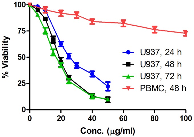
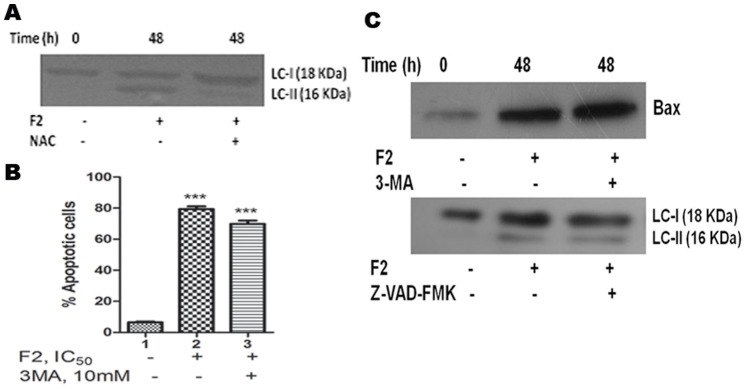
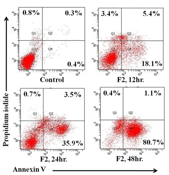
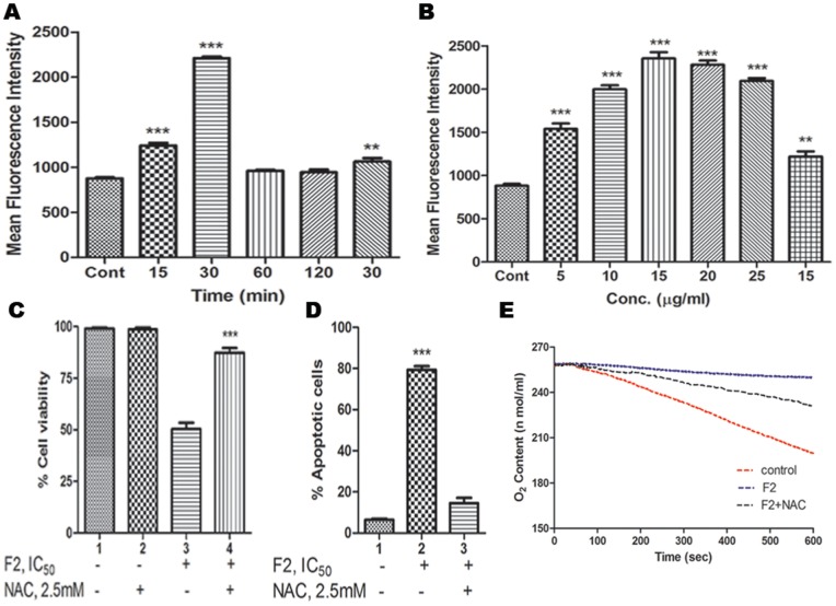
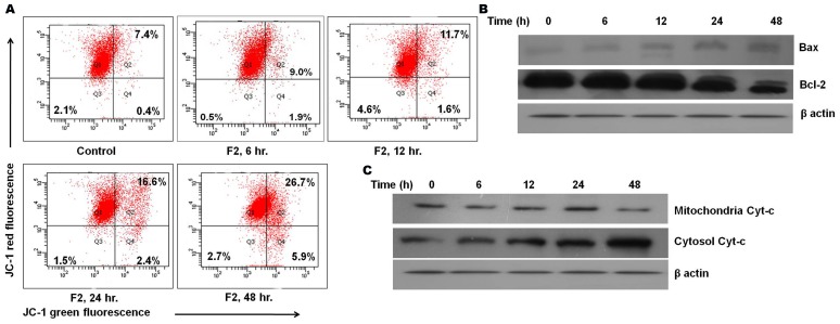
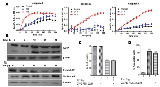
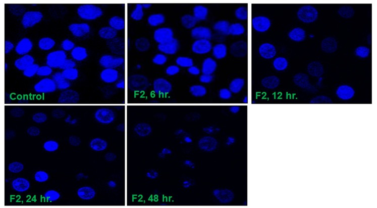
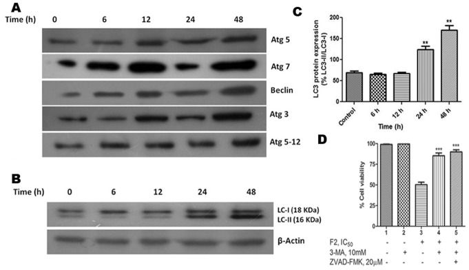
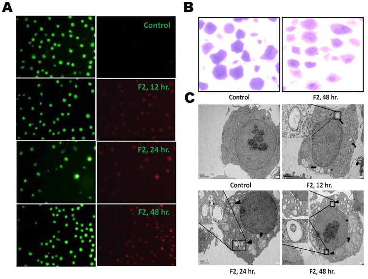
Using MTT based cell viability assay, we evaluated the cytotoxic potential of fraction F2. It was the most effective on U937 cells (IC50∶18.6 µg/ml). Inhibition of growth involved enhancement of Annexin V positivity. This was associated with elevated reactive oxygen species generation, measured by flow cytometry and reduced oxygen consumption - both effects being abrogated by anti-oxidant NAC. This caused stimulation of pro-apoptotic proteins and concomitant inhibition of anti-apoptotic protein expressions inducing mitochondrial depolarization, as measured by flow cytometry and release of cytochrome c. Interestingly, even with these molecular features of apoptosis, F2 was able to alter Atg protein levels and induce LC3 processing. This was accompanied by formation of autophagic vacuoles as revealed by fluorescence and transmission electron microscopy - confirming the occurrence of autophagy. Eventually, F2 triggered caspase cascade - executioners of programmed cell death and AIF translocation to nuclei. This culminated in cleavage of the DNA repair enzyme, poly (ADP-ribose) polymerase that caused DNA damage as proved by staining with Hoechst 33258 leading to cell death.
The findings suggest fraction F2 triggers pro-oxidant activity and mediates its cytotoxicity in leukemic cells via apoptosis and autophagy. Thus, it merits consideration and further investigation as a therapeutic option for the treatment of leukemia.
几十年来,鉴定诱导细胞凋亡的细胞毒性化合物一直是抗癌治疗的主要手段。近年来,研究重点已转移到诱导多种细胞死亡模式,同时降低全身毒性。Sesbania grandiflora 植物在印度传统医学中被广泛用于治疗多种疾病。这促使我们研究从 Sesbania grandiflora 花中分离得到的一个馏分 (F2) 在癌细胞中的抗增殖作用,并阐明其诱导细胞凋亡和自噬途径的潜在机制。
通过 MTT 基础细胞活力测定,我们评估了馏分 F2 的细胞毒性潜力。它对 U937 细胞最有效(IC50:18.6µg/ml)。生长抑制涉及 Annexin V 阳性增强。这与通过流式细胞术测量的活性氧物种生成增加以及耗氧量降低有关,这两种作用都被抗氧化剂 NAC 阻断。这导致促凋亡蛋白的刺激以及同时抑制抗凋亡蛋白表达,从而导致线粒体去极化,如通过流式细胞术和细胞色素 c 的释放所测量的。有趣的是,即使具有这些凋亡的分子特征,F2 也能够改变 Atg 蛋白水平并诱导 LC3 加工。这伴随着自噬小体的形成,如荧光和透射电子显微镜所揭示的 - 证实了自噬的发生。最终,F2 触发了 caspase 级联反应 - 程序性细胞死亡的执行者和 AIF 向细胞核的易位。这导致 DNA 修复酶多聚(ADP-核糖)聚合酶的切割,如用 Hoechst 33258 染色证明的那样导致 DNA 损伤,从而导致细胞死亡。
这些发现表明,馏分 F2 通过细胞凋亡和自噬触发促氧化剂活性,并介导其在白血病细胞中的细胞毒性。因此,它值得考虑并进一步作为白血病治疗的治疗选择进行研究。