Zhang Ping L, Mashni Joseph W, Sabbisetti Venkata S, Schworer Charles M, Wilson George D, Wolforth Stacy C, Kernen Kenneth M, Seifman Brian D, Amin Mitual B, Geddes Timothy J, Lin Fan, Bonventre Joseph V, Hafron Jason M
Department of Anatomic Pathology, William Beaumont Hospital, 3601 W. 13 Mile Rd, Royal Oak, MI, USA.
Int Urol Nephrol. 2014 Feb;46(2):379-88. doi: 10.1007/s11255-013-0522-z. Epub 2013 Aug 25.
KIM-1 staining is upregulated in proximal tubule-derived renal cell carcinoma (RCC) including clear renal cell carcinoma and papillary renal cell carcinoma, but not in chromophobe RCC (distal tubular tumor). This study was designed to prospectively examine urine KIM-1 level before and 1 month after removal of renal tumors.
A total of 19 patients were eventually enrolled in the study based on pre-operative imaging studies. Pre-operative and follow-up (1 month) urine KIM-1 levels were measured. The urine KIM-1 levels (uKIM-1) were then normalized to urine creatinine levels (uCr). Renal tumors were also stained for KIM-1 using immunohistochemical techniques.
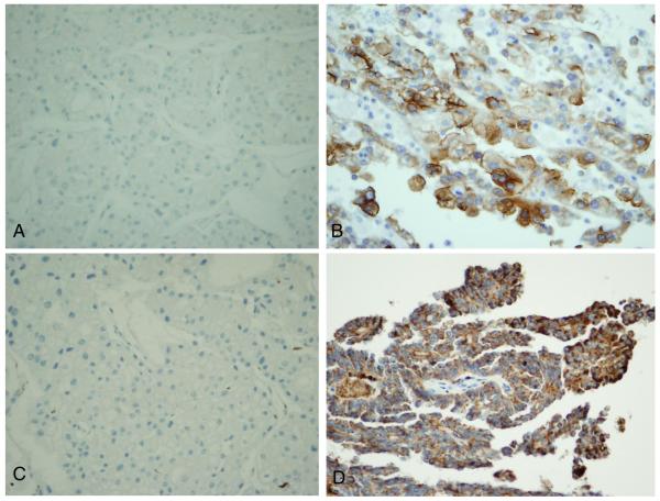
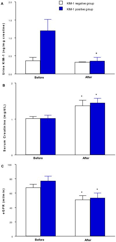
The KIM-1-negative staining group included 7 cases, and the KIM-1-positive group consisted of 12 cases. The percentage of KIM-1-positive staining RCC cells ranged from 10 to 100 %, and the staining intensity ranged from 1+ to 3+. In both groups, serum creatinine levels were both significantly elevated after nephrectomy. In the KIM-1-negative group, uKIM-1/uCr remained at a similar level before (0.37 ± 0.1 ng/mg Cr) and after nephrectomy (0.32 ± 0.01 ng/mg Cr). However, in the KIM-1-positive group, elevated uKIM-1/uCr at 1.20 ± 0.31 ng/mg Cr was significantly reduced to 0.36 ± 0.1 ng/mg Cr, which was similar to the pre-operative uKIM-1/uCr (0.37 ± 0.1 ng/mg Cr) in the KIM-1-negative group.
Our small but prospective study showed significant reduction in uKIM-1/uCr after nephrectomy in the KIM-1 positive group, suggesting that urine KIM-1 may serve as a surrogate biomarker for kidney cancer and a non-invasive pre-operative measure to evaluate the malignant potential of renal masses.
在包括透明肾细胞癌和乳头状肾细胞癌在内的近端小管来源的肾细胞癌(RCC)中,KIM-1染色上调,但在嫌色性RCC(远端小管肿瘤)中则不然。本研究旨在前瞻性地检测肾肿瘤切除术前及术后1个月的尿KIM-1水平。
基于术前影像学检查,最终共有19例患者纳入本研究。测量术前及随访(1个月)时的尿KIM-1水平。然后将尿KIM-1水平(uKIM-1)标准化为尿肌酐水平(uCr)。还采用免疫组化技术对肾肿瘤进行KIM-1染色。
KIM-1阴性染色组7例,KIM-1阳性组12例。KIM-1阳性染色的RCC细胞百分比为10%至100%,染色强度为1+至3+。两组患者肾切除术后血清肌酐水平均显著升高。在KIM-1阴性组中,肾切除术前(0.37±0.1 ng/mg Cr)和术后(0.32±0.01 ng/mg Cr)uKIM-1/uCr维持在相似水平。然而,在KIM-1阳性组中,升高的uKIM-1/uCr从1.20±0.31 ng/mg Cr显著降至0.36±0.1 ng/mg Cr,与KIM-1阴性组术前uKIM-1/uCr(0.37±0.1 ng/mg Cr)相似。
我们这项规模虽小但具有前瞻性的研究表明,KIM-1阳性组肾切除术后uKIM-1/uCr显著降低,提示尿KIM-1可能作为肾癌的替代生物标志物以及评估肾肿块恶性潜能的非侵入性术前指标。