Department of Neurosurgery, Beijing Tiantan Hospital, Capital Medical University, Beijing, China.
PLoS One. 2013 Aug 26;8(8):e72336. doi: 10.1371/journal.pone.0072336. eCollection 2013.
In order to better investigate the cause/effect relationships of human mesial temporal lobe epilepsy (mTLE), we hereby describe a new non-human primate model of mTLE.
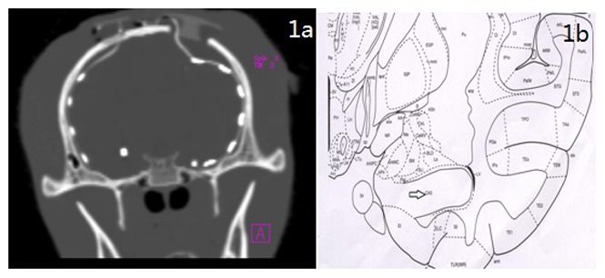
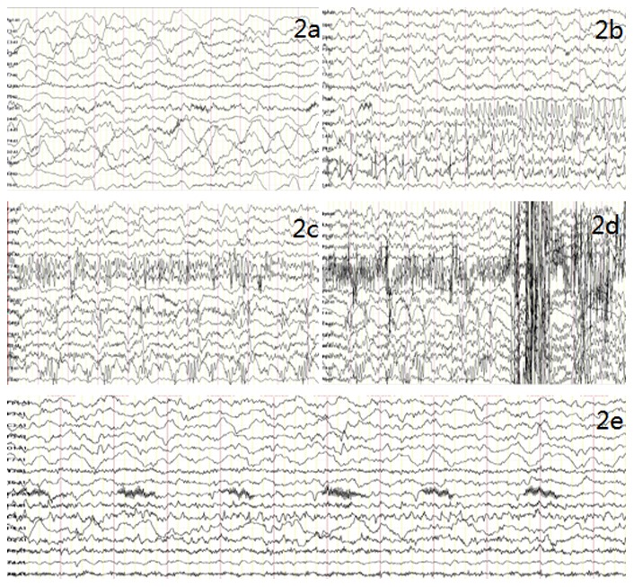
Ten macaques were studied and divided into 2 groups: saline control group (n = 4) and kainic acid (KA) injection group (n = 6). All macaques were implanted bilaterally with subdural electrodes over temporal cortex and depth electrodes in CA3 hippocampal region. KA was stereotaxically injected into the right hippocampus of macaques. All animals were monitored by video and electrocorticography (ECoG) to assess status epilepticus (SE) and subsequent spontaneous recurrent seizures (SRS). Additionally, in order to evaluate brain injury produced by SE or SRS, we used both neuroimaging, including magnetic resonance image (MRI) & magnetic resonance spectroscopy (MRS), and histological pathology, including Nissl stainning and glial fibrillary acid protein (GFAP) immunostaining.
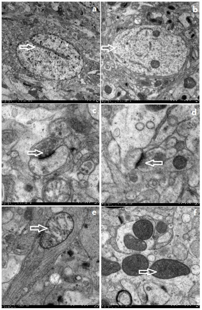
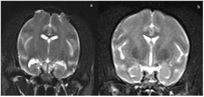
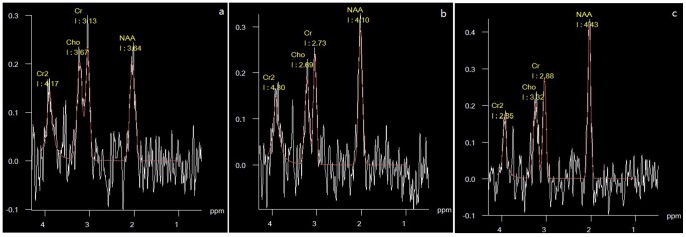
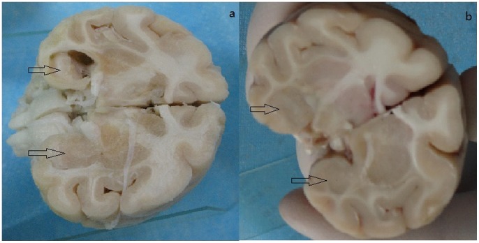
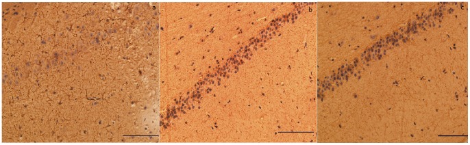
The typical seizures were observed in the KA-injected animal model. Hippocampal sclerosis could be found by MRI & MRS. Hematoxylin and eosin (H&E) staining and GFAP immunostaining showed neuronal loss, proliferation of glial cells, formation of glial scars, and hippocampal atrophy. Electron microscopic analysis of hippocampal tissues revealed neuronal pyknosis, partial ribosome depolymerization, an abnormal reduction in rough endoplasmic reticulum size, expansion of Golgi vesicles and swollen star-shaped cells. Furthermore, we reported that KA was able to induce SE followed by SRS after a variable period of time. Similar to human mTLE, brain damage is confined to the hippocampus. Accordingly, hippocampal volume is in positive correlations with the neuronal cells count in the CA3, especially the ratio of neuron/glial cell.
The results suggest that a model of mTLE can be developed in macaques by intra-hippocampal injection of KA. Brain damage is confined to the hippocampus which is similar to the human mTLE. The hippocampal volume correlates with the extension of the hippocampal damage.
为了更好地研究人类内侧颞叶癫痫(mTLE)的因果关系,我们在此描述一种新的非人类灵长类动物 mTLE 模型。
研究了 10 只猕猴,分为 2 组:生理盐水对照组(n=4)和海人酸(KA)注射组(n=6)。所有猕猴的颞叶皮质和 CA3 海马区均植入双侧硬膜下电极和深部电极。KA 立体定向注射到猕猴的右侧海马。所有动物均通过视频和脑电图(ECoG)监测以评估癫痫持续状态(SE)和随后的自发性复发性癫痫发作(SRS)。此外,为了评估 SE 或 SRS 引起的脑损伤,我们同时使用神经影像学,包括磁共振成像(MRI)和磁共振波谱(MRS),以及组织病理学,包括尼氏染色和胶质纤维酸性蛋白(GFAP)免疫染色。
在 KA 注射动物模型中观察到典型的癫痫发作。MRI 和 MRS 可发现海马硬化。苏木精和伊红(H&E)染色和 GFAP 免疫染色显示神经元丢失、胶质细胞增殖、胶质瘢痕形成和海马萎缩。海马组织的电子显微镜分析显示神经元固缩、核糖体部分解聚、粗面内质网大小异常缩小、高尔基小泡扩张和肿胀的星形细胞。此外,我们报告说 KA 能够在一段时间后诱导 SE 继而 SRS。与人类 mTLE 相似,脑损伤仅限于海马。因此,海马体积与 CA3 中的神经元细胞计数呈正相关,尤其是神经元/胶质细胞的比例。
结果表明,通过海马内注射 KA 可以在猕猴中建立 mTLE 模型。脑损伤仅限于海马,与人类 mTLE 相似。海马体积与海马损伤的扩展相关。