• 文献检索
  • 文档翻译
  • 深度研究
  • 学术资讯
  • Suppr Zotero 插件Zotero 插件
  • 邀请有礼
  • 套餐&价格
  • 历史记录
应用&插件
Suppr Zotero 插件Zotero 插件浏览器插件Mac 客户端Windows 客户端微信小程序
定价
高级版会员购买积分包购买API积分包
服务
文献检索文档翻译深度研究API 文档MCP 服务
关于我们
关于 Suppr公司介绍联系我们用户协议隐私条款
关注我们

Suppr 超能文献

核心技术专利:CN118964589B侵权必究
粤ICP备2023148730 号-1Suppr @ 2026

文献检索

告别复杂PubMed语法,用中文像聊天一样搜索,搜遍4000万医学文献。AI智能推荐,让科研检索更轻松。

立即免费搜索

文件翻译

保留排版,准确专业,支持PDF/Word/PPT等文件格式,支持 12+语言互译。

免费翻译文档

深度研究

AI帮你快速写综述,25分钟生成高质量综述,智能提取关键信息,辅助科研写作。

立即免费体验

自动化皮肤分割在乳腺癌放疗皮肤毒性的超声评估中的应用。

Automated skin segmentation in ultrasonic evaluation of skin toxicity in breast cancer radiotherapy.

机构信息

Department of Radiation Oncology, Emory University, Atlanta, Georgia, USA.

出版信息

Ultrasound Med Biol. 2013 Nov;39(11):2166-75. doi: 10.1016/j.ultrasmedbio.2013.04.006. Epub 2013 Aug 27.

DOI:10.1016/j.ultrasmedbio.2013.04.006
PMID:23993172
原文链接:https://pmc.ncbi.nlm.nih.gov/articles/PMC3913784/
Abstract

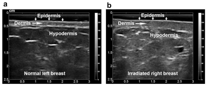
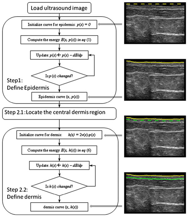
Skin toxicity is the most common side effect of breast cancer radiotherapy and impairs the quality of life of many breast cancer survivors. We, along with other researchers, have recently found quantitative ultrasound to be effective as a skin toxicity assessment tool. Although more reliable than standard clinical evaluations (visual observation and palpation), the current procedure for ultrasound-based skin toxicity measurements requires manual delineation of the skin layers (i.e., epidermis-dermis and dermis-hypodermis interfaces) on each ultrasound B-mode image. Manual skin segmentation is time consuming and subjective. Moreover, radiation-induced skin injury may decrease image contrast between the dermis and hypodermis, which increases the difficulty of delineation. Therefore, we have developed an automatic skin segmentation tool (ASST) based on the active contour model with two significant modifications: (i) The proposed algorithm introduces a novel dual-curve scheme for the double skin layer extraction, as opposed to the original single active contour method. (ii) The proposed algorithm is based on a geometric contour framework as opposed to the previous parametric algorithm. This ASST algorithm was tested on a breast cancer image database of 730 ultrasound breast images (73 ultrasound studies of 23 patients). We compared skin segmentation results obtained with the ASST with manual contours performed by two physicians. The average percentage differences in skin thickness between the ASST measurement and that of each physician were less than 5% (4.8 ± 17.8% and -3.8 ± 21.1%, respectively). In summary, we have developed an automatic skin segmentation method that ensures objective assessment of radiation-induced changes in skin thickness. Our ultrasound technology offers a unique opportunity to quantify tissue injury in a more meaningful and reproducible manner than the subjective assessments currently employed in the clinic.

摘要

皮肤毒性是乳腺癌放疗最常见的副作用,会降低许多乳腺癌幸存者的生活质量。我们与其他研究人员最近发现,定量超声是一种有效的皮肤毒性评估工具。虽然比标准临床评估(视觉观察和触诊)更可靠,但目前基于超声的皮肤毒性测量程序需要手动描绘每个超声 B 模式图像上的皮肤层(即表皮-真皮和真皮-皮下界面)。手动皮肤分割既耗时又主观。此外,辐射引起的皮肤损伤可能会降低真皮和皮下之间的图像对比度,从而增加描绘的难度。因此,我们基于主动轮廓模型开发了一种自动皮肤分割工具(ASST),并进行了两项重大修改:(i)所提出的算法引入了一种新的双曲线方案,用于双皮肤层提取,而不是原始的单主动轮廓方法。(ii)所提出的算法基于几何轮廓框架,而不是以前的参数算法。该 ASST 算法在一个包含 730 个乳腺癌超声图像(23 名患者的 73 个超声研究)的乳腺癌图像数据库上进行了测试。我们将 ASST 获得的皮肤分割结果与两名医生进行的手动轮廓进行了比较。ASST 测量的皮肤厚度与每位医生的测量结果之间的平均百分比差异小于 5%(分别为 4.8 ± 17.8%和-3.8 ± 21.1%)。总之,我们已经开发出一种自动皮肤分割方法,可确保对皮肤厚度的辐射诱导变化进行客观评估。我们的超声技术提供了一个独特的机会,可以比临床中目前使用的主观评估更有意义和可重复地量化组织损伤。

https://cdn.ncbi.nlm.nih.gov/pmc/blobs/16b2/3913784/54f250dd3f32/nihms473895f7.jpg
https://cdn.ncbi.nlm.nih.gov/pmc/blobs/16b2/3913784/b49648484232/nihms473895f1.jpg
https://cdn.ncbi.nlm.nih.gov/pmc/blobs/16b2/3913784/1d1c9de0a1de/nihms473895f2.jpg
https://cdn.ncbi.nlm.nih.gov/pmc/blobs/16b2/3913784/dfa0a1a45ae1/nihms473895f3.jpg
https://cdn.ncbi.nlm.nih.gov/pmc/blobs/16b2/3913784/fe88c3973dd2/nihms473895f4.jpg
https://cdn.ncbi.nlm.nih.gov/pmc/blobs/16b2/3913784/be9651210ddf/nihms473895f5.jpg
https://cdn.ncbi.nlm.nih.gov/pmc/blobs/16b2/3913784/8dd84ac6334a/nihms473895f6.jpg
https://cdn.ncbi.nlm.nih.gov/pmc/blobs/16b2/3913784/54f250dd3f32/nihms473895f7.jpg

相似文献

1
Automated skin segmentation in ultrasonic evaluation of skin toxicity in breast cancer radiotherapy.自动化皮肤分割在乳腺癌放疗皮肤毒性的超声评估中的应用。
Ultrasound Med Biol. 2013 Nov;39(11):2166-75. doi: 10.1016/j.ultrasmedbio.2013.04.006. Epub 2013 Aug 27.
2
Automated 3D ultrasound image segmentation to aid breast cancer image interpretation.用于辅助乳腺癌图像解读的自动化三维超声图像分割
Ultrasonics. 2016 Feb;65:51-8. doi: 10.1016/j.ultras.2015.10.023. Epub 2015 Oct 31.
3
Reliability of quantitative ultrasonic assessment of normal-tissue toxicity in breast cancer radiotherapy.定量超声评估乳腺癌放疗正常组织毒性的可靠性。
Int J Radiat Oncol Biol Phys. 2012 Feb 1;82(2):724-31. doi: 10.1016/j.ijrobp.2010.12.066. Epub 2011 Mar 23.
4
Medical breast ultrasound image segmentation by machine learning.基于机器学习的医学乳房超声图像分割。
Ultrasonics. 2019 Jan;91:1-9. doi: 10.1016/j.ultras.2018.07.006. Epub 2018 Jul 18.
5
Effect of a novel segmentation algorithm on radiologists' diagnosis of breast masses using ultrasound imaging.一种新的分割算法对超声成像中放射科医生诊断乳腺肿块的影响。
Ultrasound Med Biol. 2012 Jan;38(1):119-27. doi: 10.1016/j.ultrasmedbio.2011.09.011. Epub 2011 Nov 21.
6
A new automated method for the segmentation and characterization of breast masses on ultrasound images.一种用于超声图像上乳腺肿块分割与特征描述的新型自动化方法。
Med Phys. 2009 May;36(5):1553-65. doi: 10.1118/1.3110069.
7
Computerized lesion segmentation of breast ultrasound based on marker-controlled watershed transformation.基于标记控制分水岭变换的乳腺超声计算机病变分割。
Med Phys. 2010 Jan;37(1):82-95. doi: 10.1118/1.3265959.
8
Combining low-, high-level and empirical domain knowledge for automated segmentation of ultrasonic breast lesions.结合低层次、高层次和经验领域知识用于超声乳腺病变的自动分割
IEEE Trans Med Imaging. 2003 Feb;22(2):155-69. doi: 10.1109/TMI.2002.808364.
9
Automatic measurement of dermal thickness from B-scan ultrasound images using active contours.使用活动轮廓从B超超声图像自动测量皮肤厚度。
Skin Res Technol. 2005 May;11(2):79-90. doi: 10.1111/j.1600-0846.2005.00108.x.
10
New Fully Automated Method for Segmentation of Breast Lesions on Ultrasound Based on Texture Analysis.基于纹理分析的超声乳腺病变分割全新全自动方法
Ultrasound Med Biol. 2016 Jul;42(7):1637-50. doi: 10.1016/j.ultrasmedbio.2016.02.016. Epub 2016 Apr 16.

引用本文的文献

1
The usefulness of automated high frequency ultrasound image analysis in atopic dermatitis staging.自动高频超声图像分析在特应性皮炎分期中的应用价值。
Sci Rep. 2025 Jan 2;15(1):163. doi: 10.1038/s41598-024-84051-6.
2
Ultrasound Findings After Breast Cancer Radiation Therapy: Cutaneous, Pleural, Pulmonary, and Cardiac Changes.乳腺癌放疗后的超声表现:皮肤、胸膜、肺部和心脏改变。
Korean J Radiol. 2024 Nov;25(11):982-991. doi: 10.3348/kjr.2024.0672.
3
Computer-Aided Diagnosis Methods for High-Frequency Ultrasound Data Analysis: A Review.

本文引用的文献

1
Reliability of quantitative ultrasonic assessment of normal-tissue toxicity in breast cancer radiotherapy.定量超声评估乳腺癌放疗正常组织毒性的可靠性。
Int J Radiat Oncol Biol Phys. 2012 Feb 1;82(2):724-31. doi: 10.1016/j.ijrobp.2010.12.066. Epub 2011 Mar 23.
2
Quantitative ultrasonic evaluation of radiation-induced late tissue toxicity: pilot study of breast cancer radiotherapy.放射性迟发性组织毒性的定量超声评估:乳腺癌放疗的初步研究。
Int J Radiat Oncol Biol Phys. 2010 Nov 1;78(3):811-20. doi: 10.1016/j.ijrobp.2009.08.071. Epub 2010 Feb 19.
3
Implementation and validation of an ultrasonic tissue characterization technique for quantitative assessment of normal-tissue toxicity in radiation therapy.
基于高频超声数据分析的计算机辅助诊断方法:综述
Sensors (Basel). 2022 Oct 30;22(21):8326. doi: 10.3390/s22218326.
4
High-Frequency Ultrasound Dataset for Deep Learning-Based Image Quality Assessment.基于深度学习的图像质量评估高频超声数据集。
Sensors (Basel). 2022 Feb 14;22(4):1478. doi: 10.3390/s22041478.
5
Deep Learning-Based High-Frequency Ultrasound Skin Image Classification with Multicriteria Model Evaluation.基于深度学习的多准则模型评估的高频超声皮肤图像分类。
Sensors (Basel). 2021 Aug 30;21(17):5846. doi: 10.3390/s21175846.
6
Semi-automated weak annotation for deep neural network skin thickness measurement.基于深度学习的皮肤厚度半自动弱标注方法。
Ultrason Imaging. 2021 Jul;43(4):167-174. doi: 10.1177/01617346211014138. Epub 2021 May 11.
7
Full axillary lymph node dissection and increased breast epidermal thickness 1 year after radiation therapy for breast cancer.乳腺癌放疗 1 年后腋窝淋巴结清扫完全和乳房表皮厚度增加。
J Surg Oncol. 2019 Dec;120(8):1397-1403. doi: 10.1002/jso.25757. Epub 2019 Nov 8.
8
An Assessment of Imaging Informatics for Precision Medicine in Cancer.癌症精准医学中的影像信息学评估
Yearb Med Inform. 2017 Aug;26(1):110-119. doi: 10.15265/IY-2017-041. Epub 2017 Sep 11.
9
Objective breast tissue image classification using Quantitative Transmission ultrasound tomography.基于定量超声层析成像的乳腺组织图像分类
Sci Rep. 2016 Dec 9;6:38857. doi: 10.1038/srep38857.
10
The Impact of Axillary Lymph Node Surgery on Breast Skin Thickening During and After Radiation Therapy for Breast Cancer.腋窝淋巴结手术对乳腺癌放疗期间及放疗后乳腺皮肤增厚的影响。
Int J Radiat Oncol Biol Phys. 2016 Jun 1;95(2):590-6. doi: 10.1016/j.ijrobp.2016.01.030. Epub 2016 Jan 23.
一种用于定量评估放射治疗中正常组织毒性的超声组织表征技术的实施与验证
Med Phys. 2009 May;36(5):1643-50. doi: 10.1118/1.3103935.
4
Area and length minimizing flows for shape segmentation.用于形状分割的面积和长度最小化流。
IEEE Trans Image Process. 1998;7(3):433-43. doi: 10.1109/83.661193.
5
Speckle reducing anisotropic diffusion.斑点降噪各向异性扩散。
IEEE Trans Image Process. 2002;11(11):1260-70. doi: 10.1109/TIP.2002.804276.
6
Active contours without edges.无边缘活动轮廓。
IEEE Trans Image Process. 2001;10(2):266-77. doi: 10.1109/83.902291.
7
High frequency ultrasound assessment of skin fibrosis: clinical results.皮肤纤维化的高频超声评估:临床结果
Ultrasound Med Biol. 2007 Aug;33(8):1191-8. doi: 10.1016/j.ultrasmedbio.2007.02.009. Epub 2007 Apr 30.
8
Ultrasound image segmentation: a survey.超声图像分割:综述
IEEE Trans Med Imaging. 2006 Aug;25(8):987-1010. doi: 10.1109/tmi.2006.877092.
9
Deformable segmentation of 3-D ultrasound prostate images using statistical texture matching method.使用统计纹理匹配方法对三维超声前列腺图像进行可变形分割。
IEEE Trans Med Imaging. 2006 Mar;25(3):256-72. doi: 10.1109/TMI.2005.862744.
10
Despeckling of medical ultrasound images.医学超声图像的去斑处理
IEEE Trans Ultrason Ferroelectr Freq Control. 2006 Jan;53(1):64-78. doi: 10.1109/tuffc.2006.1588392.