• 文献检索
  • 文档翻译
  • 深度研究
  • 学术资讯
  • Suppr Zotero 插件Zotero 插件
  • 邀请有礼
  • 套餐&价格
  • 历史记录
应用&插件
Suppr Zotero 插件Zotero 插件浏览器插件Mac 客户端Windows 客户端微信小程序
定价
高级版会员购买积分包购买API积分包
服务
文献检索文档翻译深度研究API 文档MCP 服务
关于我们
关于 Suppr公司介绍联系我们用户协议隐私条款
关注我们

Suppr 超能文献

核心技术专利:CN118964589B侵权必究
粤ICP备2023148730 号-1Suppr @ 2026

文献检索

告别复杂PubMed语法,用中文像聊天一样搜索,搜遍4000万医学文献。AI智能推荐,让科研检索更轻松。

立即免费搜索

文件翻译

保留排版,准确专业,支持PDF/Word/PPT等文件格式,支持 12+语言互译。

免费翻译文档

深度研究

AI帮你快速写综述,25分钟生成高质量综述,智能提取关键信息,辅助科研写作。

立即免费体验

量子点不会改变胰腺干细胞的分化潜能,且在子细胞间随机分布。

Quantum Dots Do Not Alter the Differentiation Potential of Pancreatic Stem Cells and Are Distributed Randomly among Daughter Cells.

作者信息

Danner S, Benzin H, Vollbrandt T, Oder J, Richter A, Kruse C

机构信息

Fraunhofer Research Institution for Marine Biotechnology, 23562 Luebeck, Germany.

出版信息

Int J Cell Biol. 2013;2013:918242. doi: 10.1155/2013/918242. Epub 2013 Jul 24.

DOI:10.1155/2013/918242
PMID:23997768
原文链接:https://pmc.ncbi.nlm.nih.gov/articles/PMC3742022/
Abstract

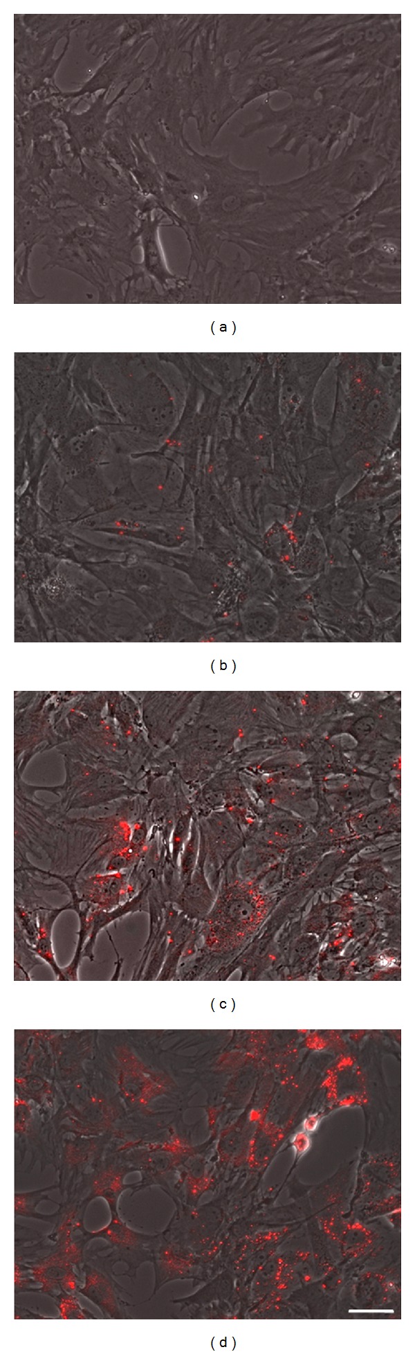
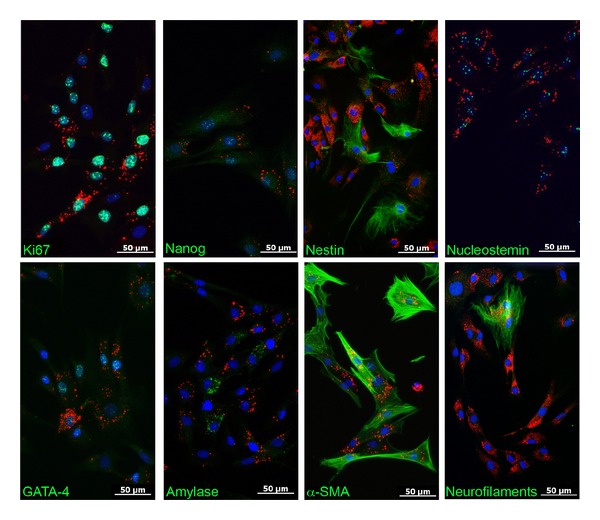
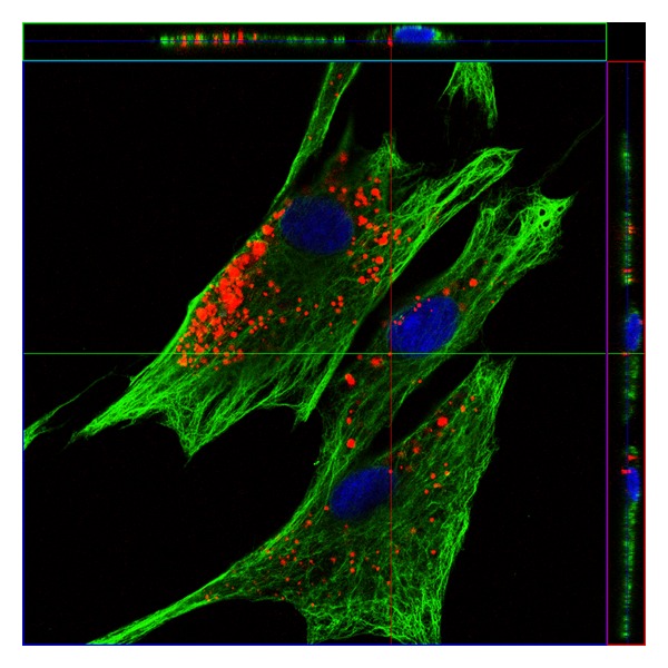
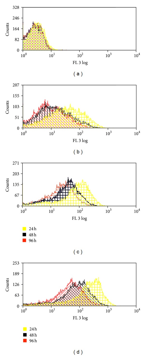
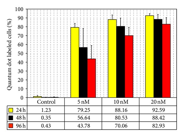
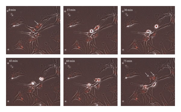
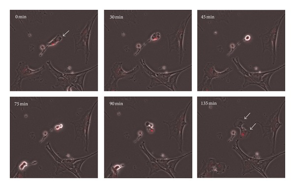
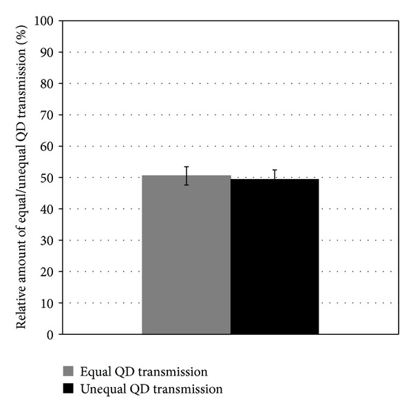
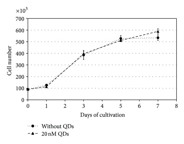
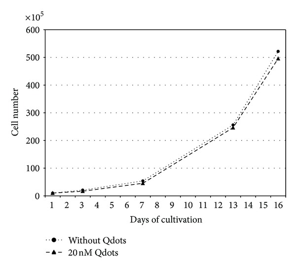
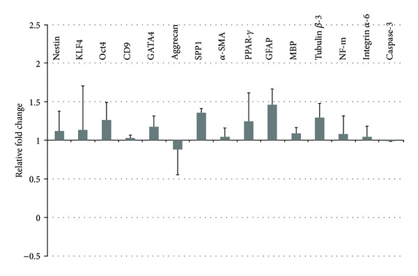
With the increasing relevance of cell-based therapies, there is a demand for cell-labeling techniques for in vitro and in vivo studies. For the reasonable tracking of transplanted stem cells in animal models, the usage of quantum dots (QDs) for sensitive cellular imaging has major advances. QDs could be delivered to the cytoplasm of the cells providing intense and stable fluorescence. Although QDs are emerging as favourable nanoparticles for bioimaging, substantial investigations are still required to consider their application for adult stem cells. Therefore, rat pancreatic stem cells (PSCs) were labeled with different concentrations of CdSe quantum dots (Qtracker 605 nanocrystals). The QD labeled PSCs showed normal proliferation and their usual spontaneous differentiation potential in vitro. The labeling of the cell population was concentration dependent, with increasing cell load from 5 nM QDs to 20 nM QDs. With time-lapse microscopy, we observed that the transmission of the QD particles during cell divisions was random, appearing as equal or unequal transmission to daughter cells. We report here that QDs offered an efficient and nontoxic way to label pancreatic stem cells without genetic modifications. In summary, QD nanocrystals are a promising tool for stem cell labeling and facilitate tracking of transplanted cells in animal models.

摘要

随着基于细胞的疗法的相关性日益增加,对用于体外和体内研究的细胞标记技术有了需求。为了在动物模型中合理追踪移植的干细胞,使用量子点(QD)进行灵敏的细胞成像有了重大进展。量子点可以递送至细胞的细胞质中,提供强烈且稳定的荧光。尽管量子点正成为用于生物成像的有利纳米颗粒,但仍需要大量研究来考虑它们在成体干细胞中的应用。因此,用不同浓度的CdSe量子点(Qtracker 605纳米晶体)标记大鼠胰腺干细胞(PSC)。量子点标记的胰腺干细胞在体外显示出正常的增殖及其通常的自发分化潜能。细胞群体的标记呈浓度依赖性,随着量子点浓度从5 nM增加到20 nM,细胞负载增加。通过延时显微镜观察,我们发现量子点颗粒在细胞分裂期间的传递是随机的,表现为向子细胞的均等或不均等传递。我们在此报告,量子点提供了一种无需基因改造即可有效且无毒地标记胰腺干细胞的方法。总之,量子点纳米晶体是一种用于干细胞标记的有前途的工具,并有助于在动物模型中追踪移植的细胞。

https://cdn.ncbi.nlm.nih.gov/pmc/blobs/b20b/3742022/347b6456ca08/IJCB2013-918242.011.jpg
https://cdn.ncbi.nlm.nih.gov/pmc/blobs/b20b/3742022/7e2e3aa1a73c/IJCB2013-918242.001.jpg
https://cdn.ncbi.nlm.nih.gov/pmc/blobs/b20b/3742022/2f77e7e2856b/IJCB2013-918242.002.jpg
https://cdn.ncbi.nlm.nih.gov/pmc/blobs/b20b/3742022/59ee849de7ff/IJCB2013-918242.003.jpg
https://cdn.ncbi.nlm.nih.gov/pmc/blobs/b20b/3742022/bd7e3430afd5/IJCB2013-918242.004.jpg
https://cdn.ncbi.nlm.nih.gov/pmc/blobs/b20b/3742022/107b000bdfbb/IJCB2013-918242.005.jpg
https://cdn.ncbi.nlm.nih.gov/pmc/blobs/b20b/3742022/cf5de39e2adb/IJCB2013-918242.006.jpg
https://cdn.ncbi.nlm.nih.gov/pmc/blobs/b20b/3742022/815b2859e023/IJCB2013-918242.007.jpg
https://cdn.ncbi.nlm.nih.gov/pmc/blobs/b20b/3742022/4e5487157a4e/IJCB2013-918242.008.jpg
https://cdn.ncbi.nlm.nih.gov/pmc/blobs/b20b/3742022/4a5841d31656/IJCB2013-918242.009.jpg
https://cdn.ncbi.nlm.nih.gov/pmc/blobs/b20b/3742022/51bda0aa935d/IJCB2013-918242.010.jpg
https://cdn.ncbi.nlm.nih.gov/pmc/blobs/b20b/3742022/347b6456ca08/IJCB2013-918242.011.jpg

相似文献

1
Quantum Dots Do Not Alter the Differentiation Potential of Pancreatic Stem Cells and Are Distributed Randomly among Daughter Cells.量子点不会改变胰腺干细胞的分化潜能,且在子细胞间随机分布。
Int J Cell Biol. 2013;2013:918242. doi: 10.1155/2013/918242. Epub 2013 Jul 24.
2
Evaluation of Human Skin-Derived Stem Cell Characteristics After Non-Invasive Quantum Dot Labeling.评价非侵入式量子点标记后人类皮肤干细胞的特性。
Cell Physiol Biochem. 2021 Jul 3;55(4):387-399. doi: 10.33594/000000391.
3
Quantum dot imaging for embryonic stem cells.用于胚胎干细胞的量子点成像。
BMC Biotechnol. 2007 Oct 9;7:67. doi: 10.1186/1472-6750-7-67.
4
Labeling of mesenchymal stem cells with bioconjugated quantum dots.用生物共轭量子点标记间充质干细胞。
Methods Mol Biol. 2011;680:61-75. doi: 10.1007/978-1-60761-901-7_4.
5
Stable and nondisruptive in vitro/in vivo labeling of mesenchymal stem cells by internalizing quantum dots.通过内化量子点实现间充质干细胞的稳定且非破坏性体外/体内标记
Hum Gene Ther. 2009 Mar;20(3):217-24. doi: 10.1089/hum.2008.100.
6
Degradation or excretion of quantum dots in mouse embryonic stem cells.量子点在小鼠胚胎干细胞中的降解或排泄。
BMC Biotechnol. 2010 May 6;10:36. doi: 10.1186/1472-6750-10-36.
7
Quantum dot labeling of mesenchymal stem cells.量子点标记间充质干细胞。
J Nanobiotechnology. 2007 Nov 7;5:9. doi: 10.1186/1477-3155-5-9.
8
Are quantum dots toxic? Exploring the discrepancy between cell culture and animal studies.量子点是否有毒?探究细胞培养与动物研究之间的差异。
Acc Chem Res. 2013 Mar 19;46(3):662-71. doi: 10.1021/ar300040z. Epub 2012 Aug 1.
9
Growth and function of equine endothelial colony forming cells labeled with semiconductor quantum dots.用半导体量子点标记的马内皮集落形成细胞的生长与功能
BMC Vet Res. 2018 Aug 23;14(1):247. doi: 10.1186/s12917-018-1572-3.
10
Lipid-Specific Labeling of Enveloped Viruses with Quantum Dots for Single-Virus Tracking.用量子点对包膜病毒进行脂质特异性标记,实现单病毒跟踪。
mBio. 2020 May 19;11(3):e00135-20. doi: 10.1128/mBio.00135-20.

引用本文的文献

1
Nanostructured scaffold as a determinant of stem cell fate.纳米结构支架作为干细胞命运的决定因素。
Stem Cell Res Ther. 2016 Dec 30;7(1):188. doi: 10.1186/s13287-016-0440-y.
2
Biodistribution and Clearance of Human Mesenchymal Stem Cells by Quantitative Three-Dimensional Cryo-Imaging After Intravenous Infusion in a Rat Lung Injury Model.大鼠肺损伤模型静脉输注后人骨髓间充质干细胞的生物分布及通过定量三维冷冻成像的清除情况
Stem Cells Transl Med. 2016 Dec;5(12):1668-1675. doi: 10.5966/sctm.2015-0379. Epub 2016 Jul 26.
3
Meta-analysis of cellular toxicity for cadmium-containing quantum dots.

本文引用的文献

1
Labeling of hematopoietic stem cells by Tat peptide conjugated quantum dots for cell tracking in mouse body.通过Tat肽偶联量子点对造血干细胞进行标记以在小鼠体内进行细胞追踪。
J Nanosci Nanotechnol. 2012 Sep;12(9):6880-6. doi: 10.1166/jnn.2012.5855.
2
Multifactorial determinants that govern nanoparticle uptake by human endothelial cells under flow.多因素决定了人内皮细胞在流动状态下对纳米颗粒的摄取。
Int J Nanomedicine. 2012;7:2943-56. doi: 10.2147/IJN.S30624. Epub 2012 Jun 14.
3
Human salivary gland stem cells: isolation, propagation, and characterization.
含镉量子点的细胞毒性的荟萃分析。
Nat Nanotechnol. 2016 May;11(5):479-86. doi: 10.1038/nnano.2015.338. Epub 2016 Feb 29.
4
In vivo tracking of human placenta derived mesenchymal stem cells in nude mice via ¹⁴C-TdR labeling.通过¹⁴C-胸腺嘧啶核苷标记在裸鼠体内追踪人胎盘来源的间充质干细胞
BMC Biotechnol. 2015 Jun 13;15:55. doi: 10.1186/s12896-015-0174-4.
5
Nanoparticle labeling identifies slow cycling human endometrial stromal cells.
Stem Cell Res Ther. 2014 Jul 4;5(4):84. doi: 10.1186/scrt473.
人唾液腺干细胞:分离、增殖及特性鉴定
Methods Mol Biol. 2012;879:403-42. doi: 10.1007/978-1-61779-815-3_25.
4
Quantum dots do not affect the behaviour of mouse embryonic stem cells and kidney stem cells and are suitable for short-term tracking.量子点不会影响小鼠胚胎干细胞和肾脏干细胞的行为,适合短期追踪。
PLoS One. 2012;7(3):e32650. doi: 10.1371/journal.pone.0032650. Epub 2012 Mar 5.
5
Cellular modulation of polymeric device surfaces: promise of adult stem cells for neuro-prosthetics.聚合物器件表面的细胞调节:成年干细胞在神经修复中的应用前景。
Front Neurosci. 2011 Oct 10;5:114. doi: 10.3389/fnins.2011.00114. eCollection 2011.
6
A novel xenogeneic co-culture system to examine neuronal differentiation capability of various adult human stem cells.一种新型的异种共培养系统,用于检测各种成人人类干细胞的神经元分化能力。
PLoS One. 2011;6(9):e24944. doi: 10.1371/journal.pone.0024944. Epub 2011 Sep 14.
7
Quantum dot labeling using positive charged peptides in human hematopoetic and mesenchymal stem cells.使用带正电荷的肽对人造血和间充质干细胞进行量子点标记。
Biomaterials. 2011 Aug;32(22):5195-205. doi: 10.1016/j.biomaterials.2011.04.004. Epub 2011 May 5.
8
Labeling of mesenchymal stem cells with bioconjugated quantum dots.用生物共轭量子点标记间充质干细胞。
Methods Mol Biol. 2011;680:61-75. doi: 10.1007/978-1-60761-901-7_4.
9
Degradation or excretion of quantum dots in mouse embryonic stem cells.量子点在小鼠胚胎干细胞中的降解或排泄。
BMC Biotechnol. 2010 May 6;10:36. doi: 10.1186/1472-6750-10-36.
10
Phenotypic indications that human sweat glands are a rich source of nestin-positive stem cell populations.表型迹象表明,人类汗腺是巢蛋白阳性干细胞群体的丰富来源。
Br J Dermatol. 2010 Feb 1;162(2):380-3. doi: 10.1111/j.1365-2133.2009.09512.x. Epub 2009 Sep 22.