Mahakian Lisa M, Farwell D Gregory, Zhang Hua, Seo Jai Woong, Poirier Brian, Tinling Steven P, Afify Alaa M, Haynam Eric M, Shaye David, Ferrara Katherine W
Department of Biomedical Engineering, University of California, Davis, 451 Health Sciences Drive, Davis, CA, 95616, USA.
Mol Imaging Biol. 2014 Apr;16(2):284-92. doi: 10.1007/s11307-013-0676-1.
Currently, 2-deoxy-2-[(18)F]fluoro-D-glucose ((18)F-FDG) is the gold standard radiotracer for staging of head and neck cancer; however, the low sensitivity of this tracer can impede detection of early lesions. (64)Cu-liposomes accumulate in various cancers and provide both a sensitive tracer and an indication of the biodistribution of nanotherapeutics. Here, the accumulation of (64)Cu-liposomes in early and established cancers is assessed and compared with (18)F-FDG in a head and neck cancer model.
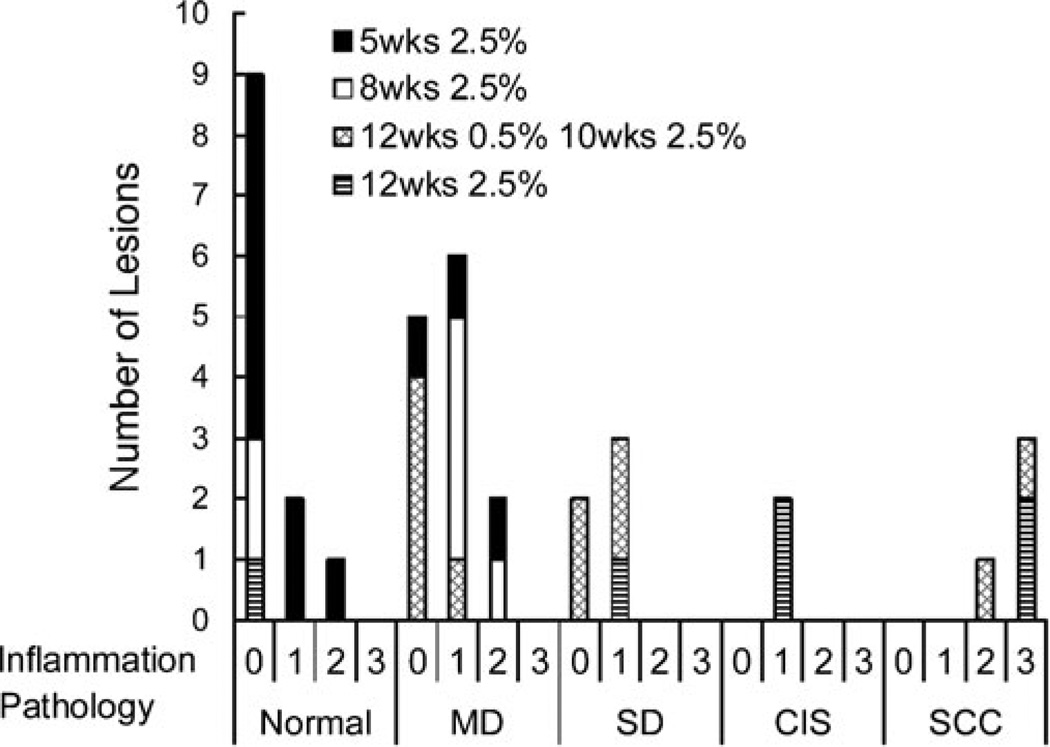
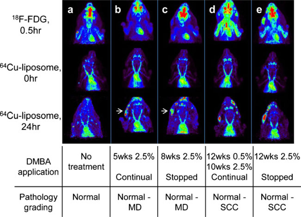
Lesions ranging from mild dysplasia to squamous cell carcinoma were induced in a hamster model of head and neck cancer by topical application of 7,12-dimethylbenz[a]anthracene to the buccal pouch. The hamsters were imaged with micro-positron emission tomography using (18)F-FDG and (64)Cu-liposomes.
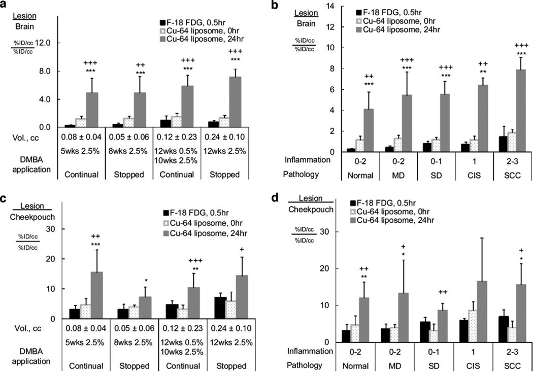
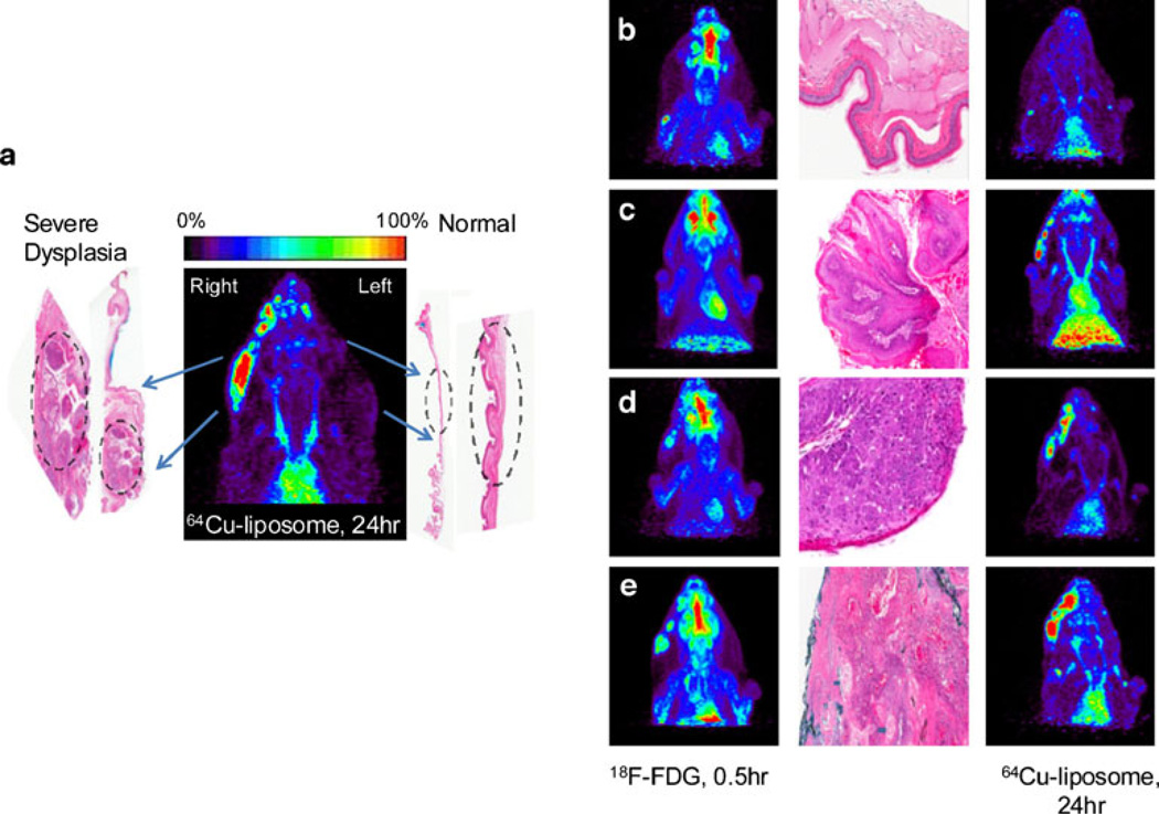
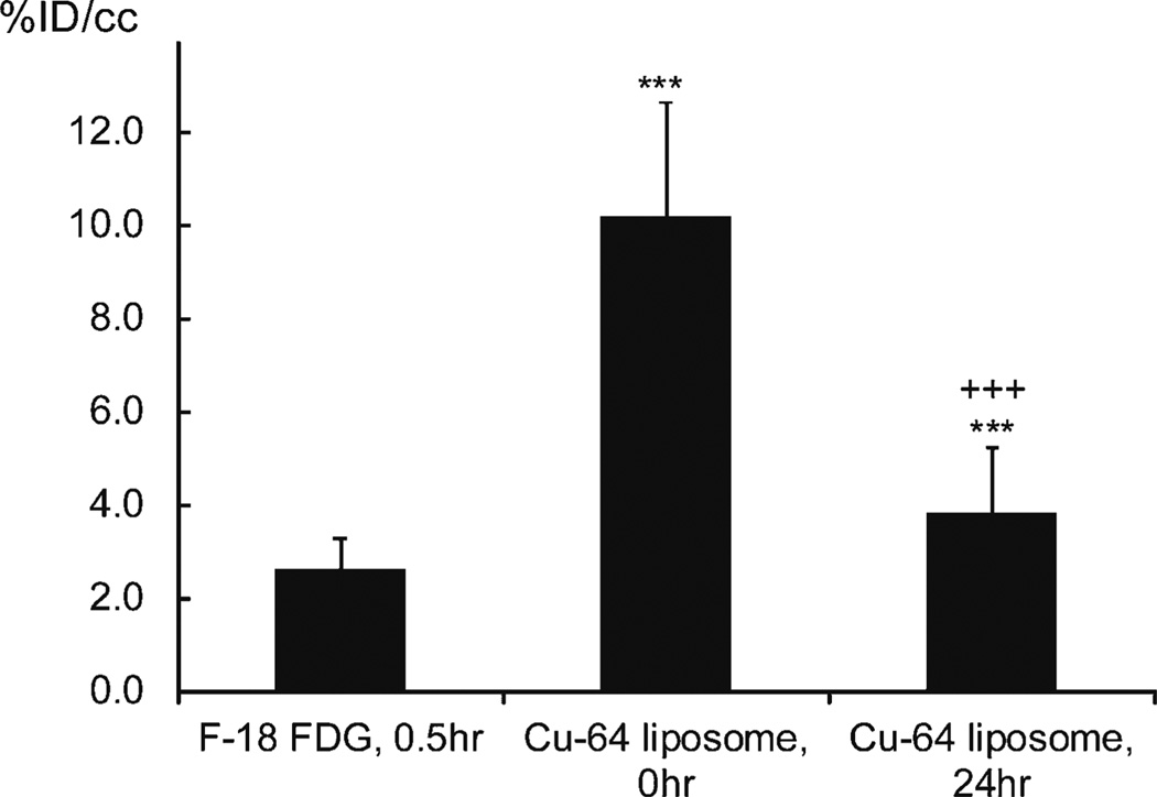
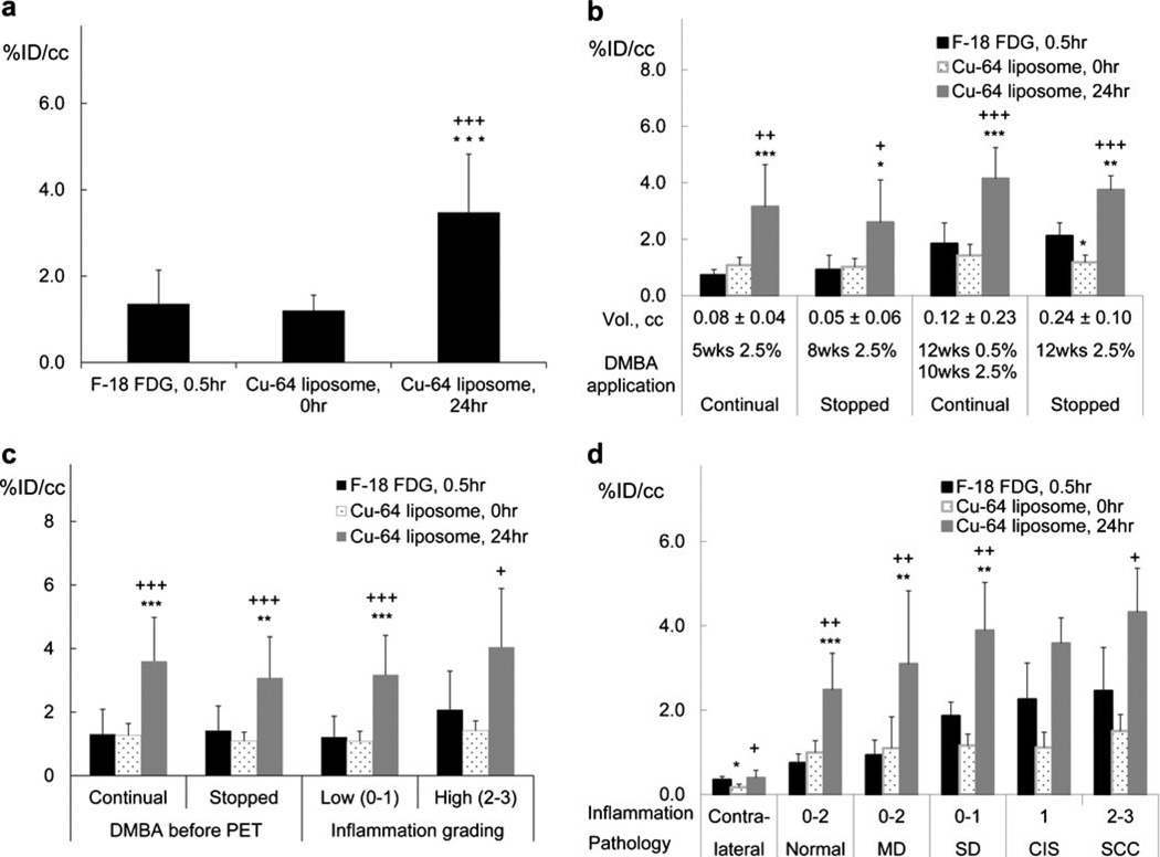
At 24 h postinjection, (64)Cu-liposome accumulation exceeded the accumulation of (18)F-FDG in every pathologic grade. The lesion-to-cheek pouch (background) ratio and lesion-to-brain ratio were also higher for (64)Cu-liposomes than for (18)F-FDG.
Imaging of a nanotracer such as (64)Cu-liposomes can improve the visualization of head and neck tumors. Accumulation of liposomal particles in head and neck tumors over various pathologic grades averaged 3.5%ID/cc demonstrating the potential for liposomal therapy with targeted chemotherapeutic agents.
目前,2-脱氧-2-[(18)F]氟-D-葡萄糖((18)F-FDG)是头颈部癌分期的金标准放射性示踪剂;然而,这种示踪剂的低灵敏度可能会妨碍早期病变的检测。(64)Cu-脂质体在各种癌症中都会蓄积,既提供了一种灵敏的示踪剂,又能显示纳米治疗药物的生物分布情况。在此,在头颈部癌模型中评估(64)Cu-脂质体在早期和已形成癌症中的蓄积情况,并与(18)F-FDG进行比较。
通过向颊囊局部应用7,12-二甲基苯并[a]蒽,在头颈部癌的仓鼠模型中诱导出从轻度发育异常到鳞状细胞癌的病变。使用(18)F-FDG和(64)Cu-脂质体对仓鼠进行微型正电子发射断层扫描成像。
注射后24小时,在每个病理分级中,(64)Cu-脂质体的蓄积量均超过(18)F-FDG的蓄积量。(64)Cu-脂质体的病变与颊囊(背景)比值和病变与脑比值也高于(18)F-FDG。
对(64)Cu-脂质体等纳米示踪剂进行成像可以改善头颈部肿瘤的可视化。脂质体颗粒在头颈部肿瘤不同病理分级中的蓄积平均为3.5%ID/cc,这表明脂质体与靶向化疗药物联合治疗具有潜力。