Hospital das Clínicas da Faculdade de Medicina da Universidade de São Paulo (FMUSP), Department of Radiology, Laboratory of Medical Investigation (LIM-44), São Paulo/SPSP, Brazil.
Clinics (Sao Paulo). 2013;68(8):1115-20. doi: 10.6061/clinics/2013(08)09.
The aim of this study was to characterize the microscopic damage to the corpus callosum in relapsing-remitting multiple sclerosis (RRMS) with diffusion tensor imaging and to investigate the correlation of this damage with disability. The diffusion tensor imaging parameters of fractional anisotropy and mean diffusivity provide information about the integrity of cell membranes, offering two more specific indices, namely the axial and radial diffusivities, which are useful for discriminating axon loss from demyelination.
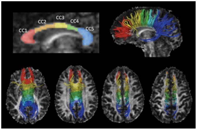
Brain magnetic resonance imaging exams of 30 relapsing-remitting multiple sclerosis patients and 30 age- and sex-matched healthy controls were acquired in a 3T scanner. The axial diffusivities, radial diffusivities, fractional anisotropy, and mean diffusivity of five segments of the corpus callosum, correlated to the Expanded Disability Status Scale score, were obtained.
All corpus callosum segments showed increased radial diffusivities and mean diffusivity, as well as decreased fractional anisotropy, in the relapsing-remitting multiple sclerosis group. The axial diffusivity was increased in the posterior midbody and splenium. The Expanded Disability Status Scale scores correlated more strongly with axial diffusivities and mean diffusivity, with an isolated correlation with radial diffusivities in the posterior midbody of the corpus callosum. There was no significant correlation with lesion loads.
Neurological dysfunction in relapsing-remitting multiple sclerosis can be influenced by commissural disconnection, and the diffusion indices of diffusion tensor imaging are potential biomarkers of disability that can be assessed during follow-up.
本研究旨在通过弥散张量成像(DTI)来描述复发缓解型多发性硬化症(RRMS)患者胼胝体的微观损伤,并探讨这种损伤与残疾的相关性。各向异性分数(FA)和平均弥散度(MD)等弥散张量成像参数可以提供细胞膜完整性的信息,而轴向弥散度和径向弥散度作为两个更具特异性的指标,有助于鉴别轴突丢失与脱髓鞘。
在 3.0T 磁共振扫描仪上采集了 30 例 RRMS 患者和 30 名年龄和性别匹配的健康对照者的脑磁共振成像检查。获得了与扩展残疾状态量表(EDSS)评分相关的胼胝体五个节段的轴向弥散度、径向弥散度、各向异性分数和平均弥散度。
RRMS 组所有胼胝体节段的径向弥散度和平均弥散度均增加,各向异性分数降低。在后体中部和压部,轴向弥散度增加。EDSS 评分与轴向弥散度和平均弥散度的相关性更强,而与后体中部的径向弥散度呈孤立相关性。与病灶负荷无明显相关性。
RRMS 患者的神经功能障碍可能受到联络纤维中断的影响,弥散张量成像的弥散指数可能是残疾的潜在生物标志物,可以在随访中进行评估。