Department of Physiology, Pharmacology, and Neuroscience, City College of the City University of New York, New York, New York, United States of America.
PLoS One. 2013 Sep 18;8(9):e74454. doi: 10.1371/journal.pone.0074454. eCollection 2013.
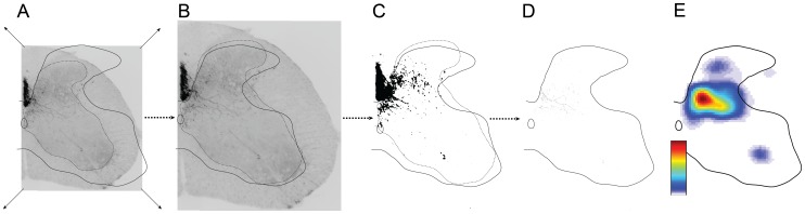
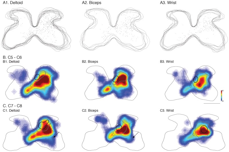
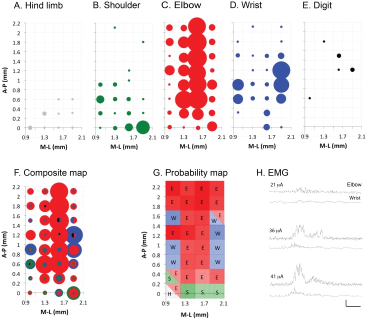
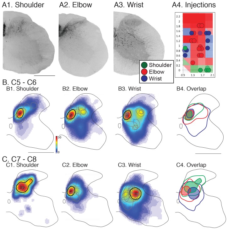
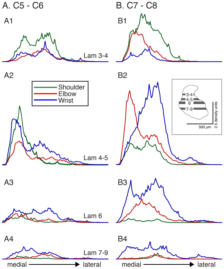
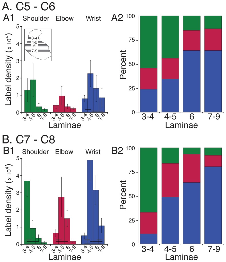
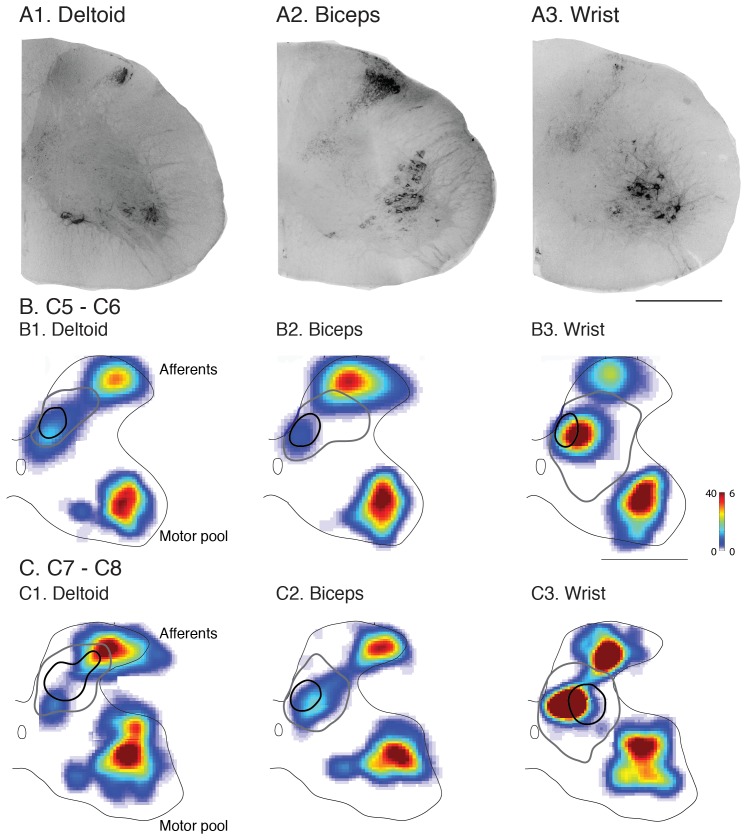
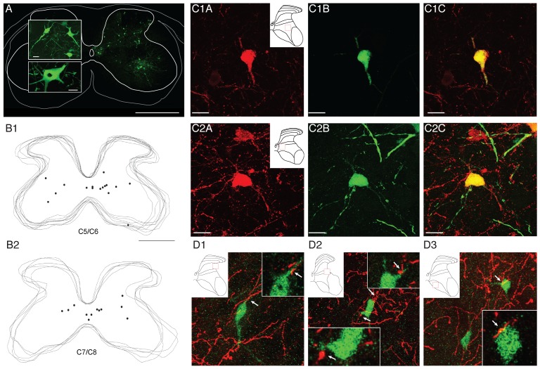
The motor cortex represents muscle and joint control and projects to spinal cord interneurons and-in many primates, including humans-motoneurons, via the corticospinal tract (CST). To examine these spinal CST anatomical mechanisms, we determined if motor cortex sites controlling individual forelimb joints project differentially to distinct cervical spinal cord territories, defined regionally and by the locations of putative last-order interneurons that were transneuronally labeled by intramuscular injection of pseudorabies virus. Motor cortex joint-specific sites were identified using intracortical-microstimulation. CST segmental termination fields from joint-specific sites, determined using anterograde tracers, comprised a high density core of terminations that was consistent between animals and a surrounding lower density projection that was more variable. Core terminations from shoulder, elbow, and wrist control sites overlapped in the medial dorsal horn and intermediate zone at C5/C6 but were separated at C7/C8. Shoulder sites preferentially terminated dorsally, in the dorsal horn; wrist/digit sites, more ventrally in the intermediate zone; and elbow sites, medially in the dorsal horn and intermediate zone. Pseudorabies virus injected in shoulder, elbow, or wrist muscles labeled overlapping populations of predominantly muscle-specific putative premotor interneurons, at a survival time for disynaptic transfer from muscle. At C5/C6, CST core projections from all joint zones were located medial to regions of densely labeled last-order interneurons, irrespective of injected muscle. At C7/C8 wrist CST core projections overlapped the densest interneuron territory, which was located in the lateral intermediate zone. In contrast, elbow CST core projections were located medial to the densest interneuron territories, and shoulder CST core projections were located dorsally and only partially overlapped the densest interneuron territory. Our findings show a surprising fractionation of CST terminations in the caudal cervical enlargement that may be organized to engage different spinal premotor circuits for distal and proximal joint control.
运动皮层代表肌肉和关节控制,并通过皮质脊髓束 (CST) 投射到脊髓中间神经元和 - 在许多灵长类动物,包括人类 - 运动神经元。为了研究这些脊髓 CST 解剖学机制,我们确定了控制单个前肢关节的运动皮层部位是否会以不同的方式投射到不同的颈椎脊髓区域,这些区域通过肌肉内注射伪狂犬病病毒进行顺行示踪,根据假定的最后一级中间神经元的位置和位置来区域性定义。使用皮层内微刺激来确定运动皮层关节特异性部位。使用顺行示踪剂确定的来自关节特异性部位的 CST 节段终止场,包括动物之间一致的高密度终止核心和更可变的周围低密度投影。来自肩部、肘部和腕部控制部位的核心终止在 C5/C6 的背角和中间区域重叠,但在 C7/C8 处分离。肩部部位优先在背角的背侧终止;腕部/指部部位更多地在中间区域的腹侧终止;肘部部位更多地在背角和中间区域的内侧终止。在肩部、肘部或腕部肌肉中注射伪狂犬病病毒标记了重叠的主要是肌肉特异性假定的运动前神经元群体,在从肌肉进行双突触传递的存活时间内。在 C5/C6,来自所有关节区的 CST 核心投射位于密集标记的最后一级中间神经元区域的内侧,无论注射的肌肉如何。在 C7/C8 腕 CST 核心投射与最密集的中间神经元区域重叠,该区域位于外侧中间区域。相比之下,肘部 CST 核心投射位于最密集的中间神经元区域的内侧,肩部 CST 核心投射位于背侧,仅部分重叠最密集的中间神经元区域。我们的发现显示 CST 终止在尾侧颈膨大中的惊人细分,这可能组织起来以用于远端和近端关节控制的不同脊髓运动前回路。