Aljohani Ayman J, Munguba Gustavo C, Guerra Yenifer, Lee Richard K, Bhattacharya Sanjoy K
Bascom Palmer Eye Institute, University of Miami, Miami, FL.
Mol Vis. 2013 Sep 19;19:1966-84. eCollection 2013.
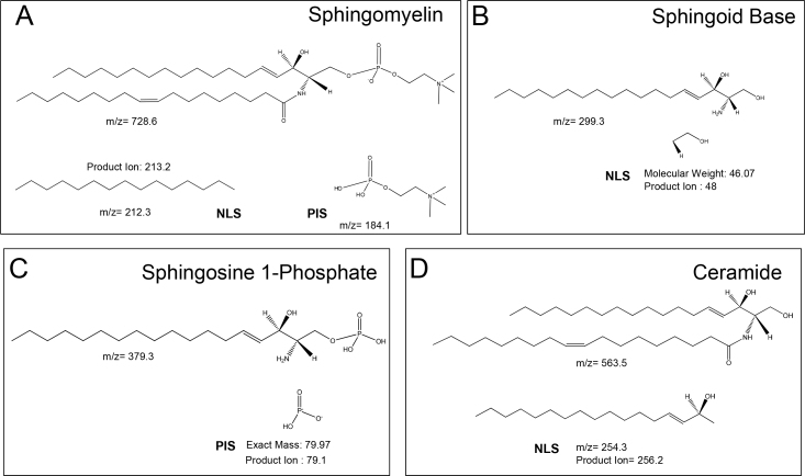
To determine the differential profiles of sphingomyelin, sphingoid base, sphingoid base-1-phosphate and ceramide lipid species and their quantitative differences between control and glaucomatous aqueous humor (AQH) derived from human donors.
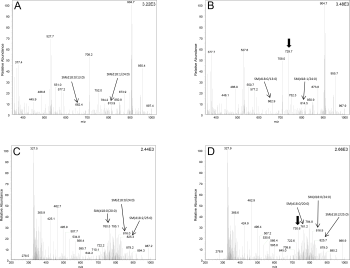
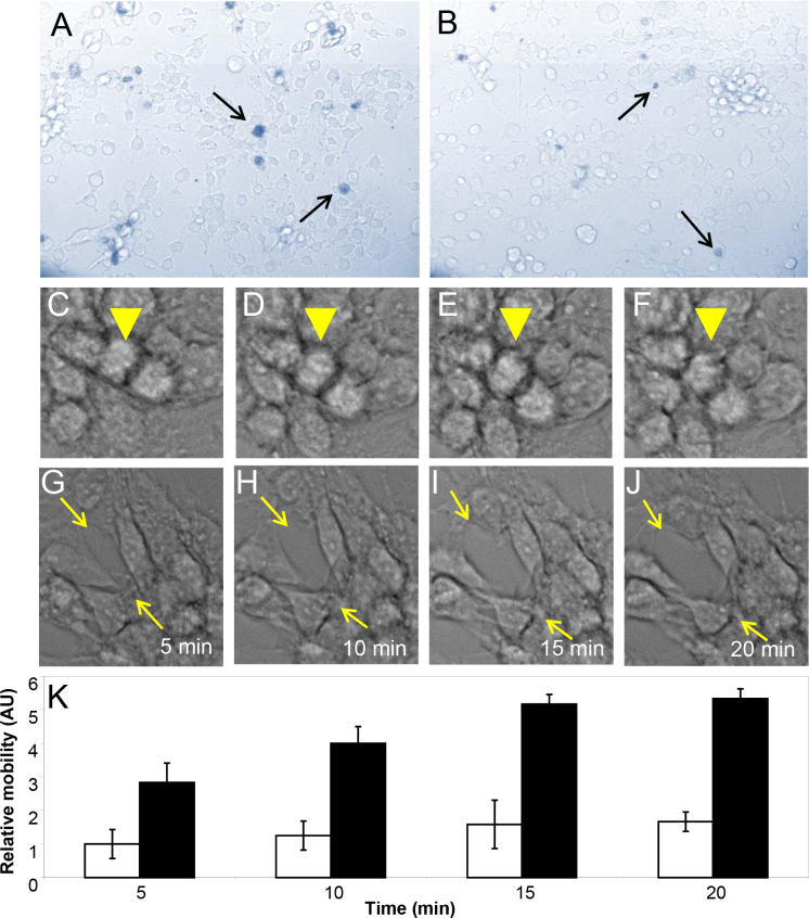
AQH from control and primary open-angle glaucoma donors was collected and subjected to lipid extraction using suitable modifications of the Bligh and Dyer method. Proteins were estimated using Bradford's method. Lipids were identified and ratiometrically quantified in a two-step process using precursor ion scan or neutral loss scan (NLS) with appropriate class-specific lipid standards on a TSQ Quantum Access Max mass spectrometer following established procedures. Primary human trabecular meshwork cells and video microscopic imaging were used to assess changes in cell shape and motility upon exposure to 20 pmol of Cer(d18:0/18:1(9Z)) in 10% dimethyl sulfoxide (vehicle).
We identified several species of sphingomyelin, sphingoid base, sphingoid base-1-phosphate, and ceramides that were common between control and glaucomatous AQH. Some unique lipid species in these classes were also identified in controls but not in glaucoma and vice versa. We found exposure to 20 pmol of Cer(d18:0/18:1(9Z)) resulted in changes in the trabecular meshwork cell shape and observed motility changes compared to vehicle-only control.
Most lipids belonging to the sphingomyelin, sphingoid base, sphingoid base-1-phosphate, and ceramide species were common between control and primary open-angle glaucoma donors. However, some sphingolipids and ceramides were found to be uniquely present in control but absent in the glaucomatous AQH and vice versa. Identification of unique lipid species present or absent in the pathophysiological context may contribute further insight into glaucoma pathology.
确定鞘磷脂、鞘氨醇碱、鞘氨醇碱-1-磷酸和神经酰胺脂质种类的差异谱,以及来自人类供体的对照和青光眼房水(AQH)之间的定量差异。
收集对照和原发性开角型青光眼供体的房水,并使用布莱和戴尔方法的适当改良进行脂质提取。使用布拉德福德法估算蛋白质含量。脂质在两步过程中通过前体离子扫描或中性丢失扫描(NLS)进行鉴定和比例定量,在TSQ Quantum Access Max质谱仪上按照既定程序使用适当的类别特异性脂质标准品。使用原代人小梁网细胞和视频显微镜成像来评估在10%二甲基亚砜(载体)中暴露于20 pmol Cer(d18:0/18:1(9Z))后细胞形状和运动性的变化。
我们鉴定出对照和青光眼房水中常见的几种鞘磷脂、鞘氨醇碱、鞘氨醇碱-1-磷酸和神经酰胺。在这些类别中,一些独特的脂质种类在对照中也被鉴定出,但在青光眼中未被鉴定出,反之亦然。我们发现暴露于20 pmol Cer(d18:0/18:1(9Z))会导致小梁网细胞形状发生变化,并观察到与仅使用载体的对照相比运动性发生了变化。
对照和原发性开角型青光眼供体中,大多数属于鞘磷脂、鞘氨醇碱、鞘氨醇碱-1-磷酸和神经酰胺种类的脂质是常见的。然而,发现一些鞘脂和神经酰胺仅在对照中存在,而在青光眼房水中不存在,反之亦然。鉴定在病理生理背景下存在或不存在的独特脂质种类可能有助于进一步深入了解青光眼病理。