Departamento de Genética, Universidade do Estado do Rio de Janeiro, Instituto de Biologia Roberto Alcantara Gomes, 20550-013 Rio de Janeiro, Brazil.
Radiat Oncol. 2013 Oct 5;8:231. doi: 10.1186/1748-717X-8-231.
MicroRNAs are non-coding RNAs involved in the regulation of gene expression including DNA damage responses. Low doses of low energy X-ray radiation, similar to those used in mammographic exams, has been described to be genotoxic. In the present work we investigated the expression of miR-34a; a well described p53-regulated miRNA implicated in cell responses to X-ray irradiation at low doses.
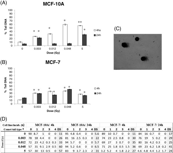
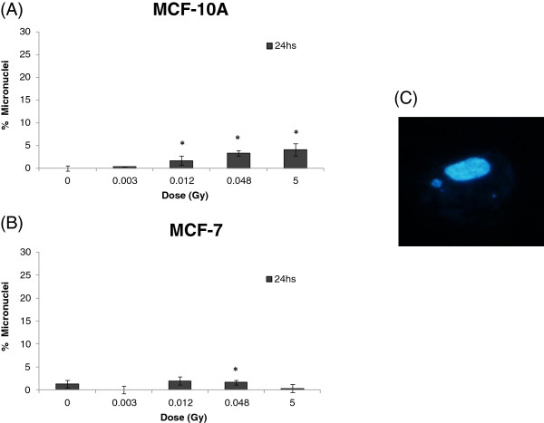
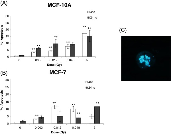
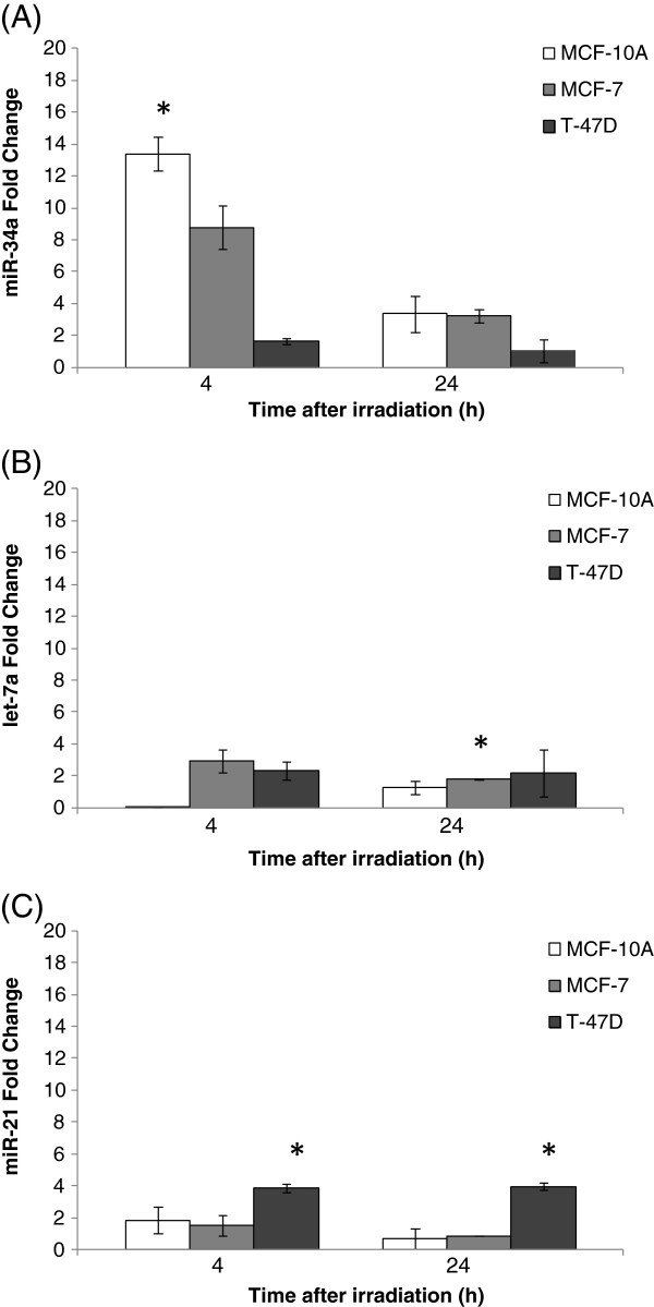
Non-cancerous breast cell line MCF-10A and cancerous T-47D and MCF-7 cell lines were submitted to a low-energy X-ray irradiation (ranging from 28-30 Kv) using a dose of 5 Gy. The expression level of miR-34a, let-7a and miR-21 was assessed by qRT-PCR at 4 and 24 hours post-irradiation. DNA damage was then measured by comet assay and micronuclei estimation in MCF-10A and MCF-7 cell lines, where an increase of miR-34a levels could be observed after irradiation. The rate of apoptotic cells was estimated by nuclear staining and fluorescence microscopy. These experiments were also performed at low doses (3; 12 and 48 mGy) in MCF-10A and MCF-7 cell lines.
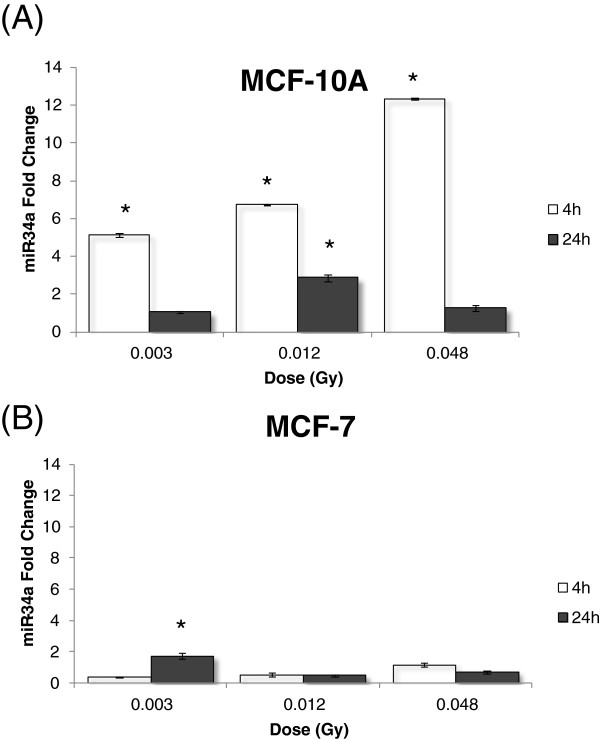
We have observed an increase in miR-34a expression 4 hours post-irradiation at 5 Gy in MCF-10A and MCF-7 cell lines while its level did not change in T-47D, a breast cancer cell line bearing non-functional p53. At low doses, miR-34a was up-regulated in non-tumoral MCF-10A to a higher extent as compared to MCF-7. MiR-34a levels decreased 24 hours post-irradiation. We have also observed DNA damage and apoptosis at low-energy X-ray irradiation at low doses and the high dose in MCF-10A and MCF-7 4 and 24 hours post-irradiation relative to the mock control.
Low energy X-ray is able to promote DNA strand breaks and miR-34a might be involved in cell responses to low energy X-ray DNA damage. MiR-34a expression correlates with X-ray dose, time after irradiation and cell type. The present study reinforces the need of investigating consequences of low dose X-ray irradiation of breast cells.
MicroRNAs 是参与基因表达调控的非编码 RNA,包括 DNA 损伤反应。类似于乳房 X 光检查中使用的低能量 X 射线辐射,已被描述为具有遗传毒性。在本工作中,我们研究了 miR-34a 的表达; 一种已被描述的 p53 调控 miRNA,与低剂量 X 射线照射的细胞反应有关。
非癌细胞系 MCF-10A 和癌细胞系 T-47D 和 MCF-7 细胞系用低能量 X 射线(范围从 28-30 Kv)照射,剂量为 5 Gy。在照射后 4 小时和 24 小时,通过 qRT-PCR 评估 miR-34a、let-7a 和 miR-21 的表达水平。然后通过彗星试验和微核估计测量 MCF-10A 和 MCF-7 细胞系中的 DNA 损伤,其中照射后可以观察到 miR-34a 水平的增加。通过核染色和荧光显微镜估计凋亡细胞的比率。这些实验也在 MCF-10A 和 MCF-7 细胞系中低剂量(3、12 和 48 mGy)下进行。
我们观察到 MCF-10A 和 MCF-7 细胞系在 5 Gy 照射后 4 小时 miR-34a 表达增加,而在乳腺癌细胞系 T-47D 中其水平没有变化,T-47D 携带无功能的 p53。在低剂量下,非肿瘤 MCF-10A 中 miR-34a 的上调程度高于 MCF-7。照射后 24 小时 miR-34a 水平下降。我们还观察到 MCF-10A 和 MCF-7 在低能量 X 射线照射下的 DNA 损伤和细胞凋亡,与假对照相比,在照射后 4 小时和 24 小时,剂量相对较高。
低能 X 射线能够促进 DNA 链断裂,miR-34a 可能参与细胞对低能 X 射线 DNA 损伤的反应。miR-34a 的表达与 X 射线剂量、照射后时间和细胞类型有关。本研究加强了对乳腺细胞低剂量 X 射线照射后果进行研究的必要性。