Lu Kai-hua, Li Wei, Liu Xiang-hua, Sun Ming, Zhang Mei-ling, Wu Wei-qin, Xie Wei-ping, Hou Ya-yi
Department of respiratory, First Affiliated Hospital, Nanjing Medical University, Nanjing, People's Republic of China.
BMC Cancer. 2013 Oct 7;13:461. doi: 10.1186/1471-2407-13-461.
Long non-coding RNAs play an important role in tumorigenesis, hence, identification of cancer-associated lncRNAs and investigation of their biological functions and molecular mechanisms are important for understanding the development and progression of cancer. Recently, the downregulation of lncRNA MEG3 has been observed in various human cancers. However, its role in non-small cell lung cancer (NSCLC) is unknown. The aim of this study was to examine the expression pattern of MEG3 in NSCLC and to evaluate its biological role and clinical significance in tumor progression.
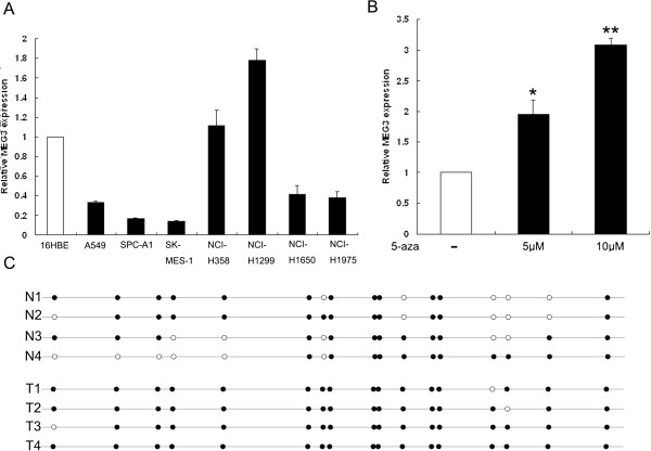
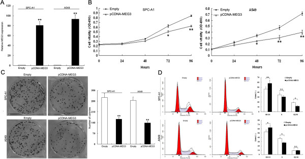
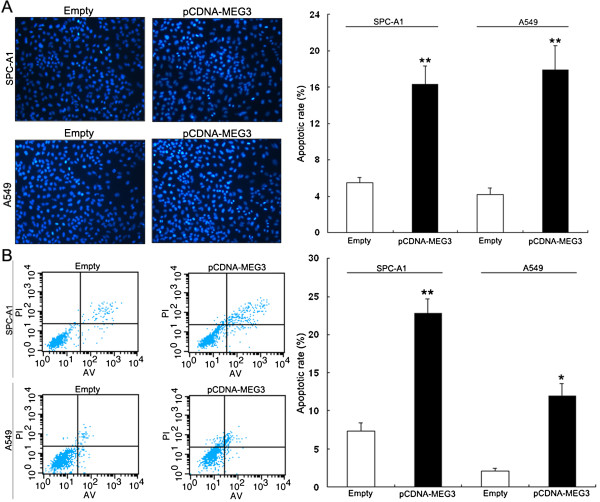
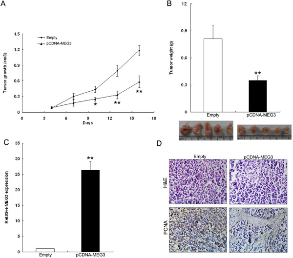
Expression of MEG3 was analyzed in 44 NSCLC tissues and 7 NSCLC cell lines by qRT-PCR. Over-expression approaches were used to investigate the biological functions of MEG3 in NSCLC cells. Bisulfite sequencing was used to investigate DNA methylation on MEG3 expression. The effect of MEG3 on proliferation was evaluated by MTT and colony formation assays, and cell apoptosis was evaluated by Hoechst staining and Flow-cytometric analysis. NSCLC cells transfected with pCDNA-MEG3 were injection into nude mice to study the effect of MEG3 on tumorigenesis in vivo . Protein levels of MEG3 targets were determined by western blot analysis. Differences between groups were tested for significance using Student's t-test (two-tailed).
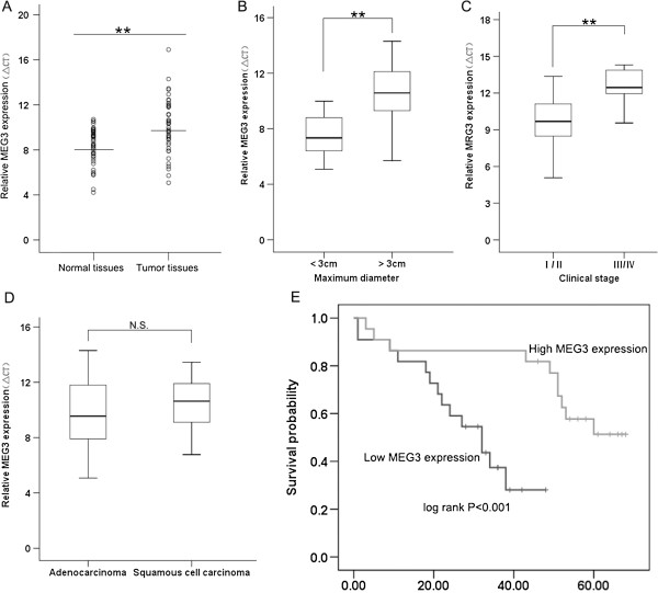
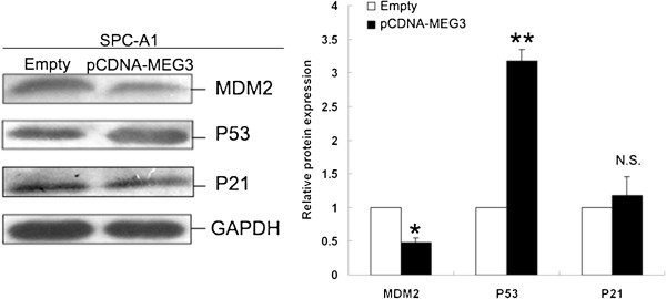
MEG3 expression was decreased in non-small cell lung cancer (NSCLC) tumor tissues compared with normal tissues, and associated with advanced pathologic stage, and tumor size. Moreover, patients with lower levels of MEG3 expression had a relatively poor prognosis. Overexpression of MEG3 decreased NSCLC cells proliferation and induced apoptosis in vitro and impeded tumorigenesis in vivo. MDM2 and p53 protein levels were affected by MEG3 over-expression in vitro.
Our findings indicate that MEG3 is significantly down-regulated in NSCLC tissues that could be affected by DNA methylation, and regulates NSCLC cell proliferation and apoptosis, partially via the activition of p53. Thus, MEG3 may represent a new marker of poor prognosis and is a potential therapeutic target for NSCLC intervention.
长链非编码RNA在肿瘤发生中起重要作用,因此,鉴定与癌症相关的长链非编码RNA并研究其生物学功能和分子机制对于理解癌症的发生和发展至关重要。最近,在各种人类癌症中均观察到lncRNA MEG3的表达下调。然而,其在非小细胞肺癌(NSCLC)中的作用尚不清楚。本研究的目的是检测MEG3在NSCLC中的表达模式,并评估其在肿瘤进展中的生物学作用和临床意义。
采用qRT-PCR分析44例NSCLC组织和7株NSCLC细胞系中MEG3的表达。采用过表达方法研究MEG3在NSCLC细胞中的生物学功能。采用亚硫酸氢盐测序法研究MEG3表达的DNA甲基化情况。通过MTT和集落形成试验评估MEG3对增殖的影响,通过Hoechst染色和流式细胞术分析评估细胞凋亡。将转染pCDNA-MEG3的NSCLC细胞注射到裸鼠体内,研究MEG3对体内肿瘤发生的影响。通过蛋白质免疫印迹分析确定MEG3靶点的蛋白质水平。使用Student's t检验(双侧)检测组间差异的显著性。
与正常组织相比,非小细胞肺癌(NSCLC)肿瘤组织中MEG3表达降低,且与病理分期较晚和肿瘤大小相关。此外,MEG3表达水平较低的患者预后相对较差。MEG3过表达在体外降低了NSCLC细胞的增殖并诱导了细胞凋亡,在体内抑制了肿瘤发生。体外MEG3过表达影响了MDM2和p53蛋白水平。
我们的研究结果表明,MEG3在NSCLC组织中显著下调,可能受DNA甲基化影响,并部分通过激活p53调节NSCLC细胞的增殖和凋亡。因此,MEG3可能是预后不良的新标志物,是NSCLC干预的潜在治疗靶点。