Center Laboratory of Stomatology, Stomatological Hospital Affiliated Medical School, Nanjing University, Nanjing, People's Republic of China.
Int J Nanomedicine. 2013;8:3737-44. doi: 10.2147/IJN.S52135. Epub 2013 Oct 7.
The successful biotherapy of carcinoma with dendritic cell (DC) vaccines pivotally relies on DCs' migratory capability into lymph tissues and activation of T cells. Accurate imaging and evaluation of DC migration in vivo have great significance during antitumor treatment with DC vaccine. We herein examined the behavior of DCs influenced by synthetic superparamagnetic iron oxide (SPIO) nanoparticle labeling.
γ-Fe2O3 nanoparticles were prepared and DCs, which were induced from bone marrow monocytes of enhanced green fluorescent protein (EGFP) transgenic mice, were labeled. The endocytosis of the SPIO, surface molecules, cell apoptosis and fluorescence intensity of EGFP-DCs were displayed by Prussian blue staining and flow cytometry (FCM), respectively. After EGFP-DCs, labeled with SPIO, were injected into footpads (n = 5) for 24 hours, the mice were examined in vivo by optical imaging (OPI). Meanwhile, confocal imaging and FCM were applied, respectively, to detect the migration of labeled DCs into draining lymph nodes.
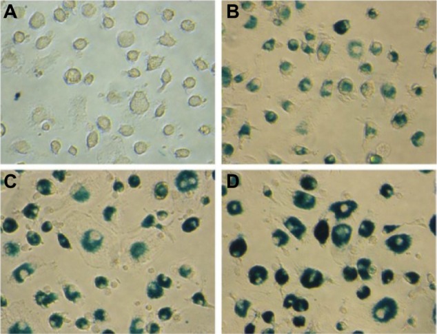
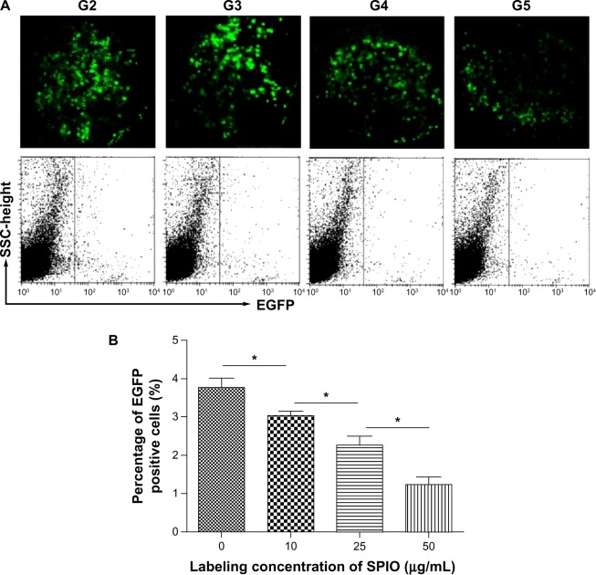
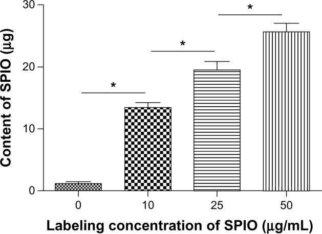
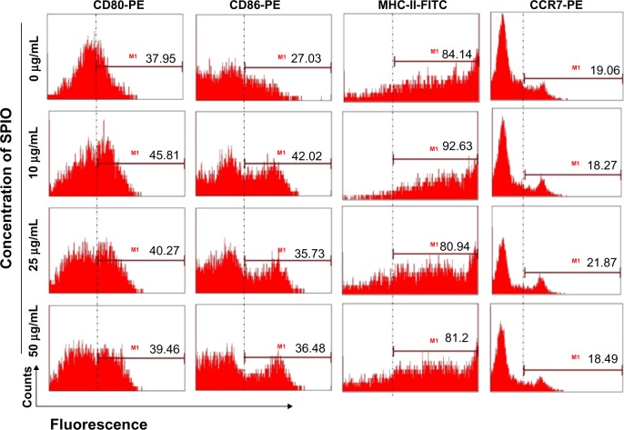
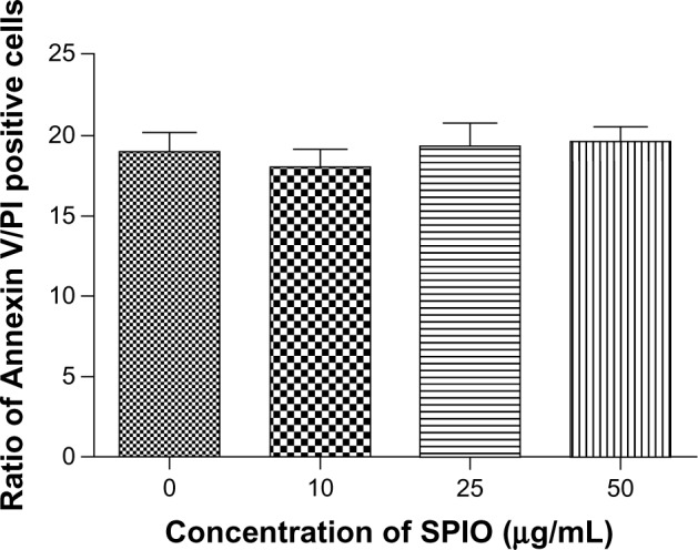
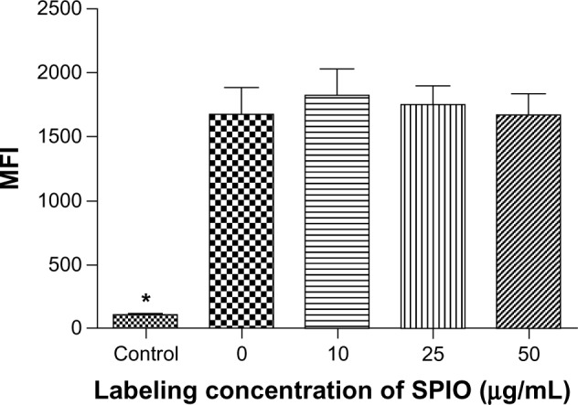
Nearly 100% of cells were labeled by the SPIO, in which the intracellular blue color gradually deepened and the iron contents rose with the increase of labeling iron concentrations. In addition, cell apoptosis and the surface molecules on DCs were at similar levels after SPIO labeling. After confirming that the fluorescence intensity of EGFP on DCs was not influenced by SPIO, the homing ability of EGFP-DCs labeled with SPIO displayed that the fluorescence intensity and the ratios of EGFP-DCs in draining lymph nodes were gradually decreased with the increase of labeling iron concentrations.
The synthetic SPIO nanoparticles possess perfect labeling ability and biocompatibility. Moreover, DCs labeled with a low dose of SPIO showed stronger migratory capability in vivo.
树突状细胞(DC)疫苗的成功的生物治疗主要依赖于 DC 向淋巴组织的迁移能力和 T 细胞的激活。在使用 DC 疫苗进行抗肿瘤治疗时,准确地对 DC 迁移进行成像和评估具有重要意义。我们在此检查了合成超顺磁性氧化铁(SPIO)纳米颗粒标记对 DC 行为的影响。
制备γ-Fe2O3 纳米颗粒,并对来自增强型绿色荧光蛋白(EGFP)转基因小鼠骨髓单核细胞诱导的 DC 进行标记。通过普鲁士蓝染色和流式细胞术(FCM)分别显示 SPIO 的内吞作用、表面分子、细胞凋亡和 EGFP-DC 的荧光强度。将 SPIO 标记的 EGFP-DC 注入脚掌(n = 5)24 小时后,通过光学成像(OPI)对小鼠进行体内检查。同时,分别应用共聚焦成像和 FCM 检测标记的 DC 向引流淋巴结的迁移。
几乎 100%的细胞被 SPIO 标记,其中细胞内蓝色逐渐加深,铁含量随着标记铁浓度的增加而升高。此外,SPIO 标记后 DC 上的细胞凋亡和表面分子水平相似。在确认 SPIO 对 EGFP-DC 的荧光强度没有影响后,SPIO 标记的 EGFP-DC 的归巢能力显示,随着标记铁浓度的增加,荧光强度和引流淋巴结中 EGFP-DC 的比例逐渐降低。
合成的 SPIO 纳米颗粒具有完美的标记能力和生物相容性。此外,用低剂量 SPIO 标记的 DC 显示出更强的体内迁移能力。