1. Department of Medicine, Division of Gastroenterology, Hepatology and Nutrition, University of Pittsburgh, Pittsburgh, Pennsylvania, USA;
J Cancer. 2013 Sep 10;4(8):626-34. doi: 10.7150/jca.6990. eCollection 2013.
Barrett's esophagus (BE) affects up to 12 million Americans and confers an increased risk for development of esophageal adenocarcinoma (EAC). EAC is often fatal unless detected early. Given the high prevalence, high cost of surveillance and relatively low risk of most affected individuals, identification of high-risk patients for additional scrutiny, regular surveillance, or ablative therapy is crucial. The exploration of "field effect" by probing uninvolved esophageal mucosa to predict the risk of EAC has the potential as an improved surveillance and prevention strategy. In this study, we evaluate the ability of nuclear nano-architecture markers from normal squamous esophagus and gastric cardia to detect the "field effect" of esophageal dysplasia and EAC, and their response to endoscopic therapy.
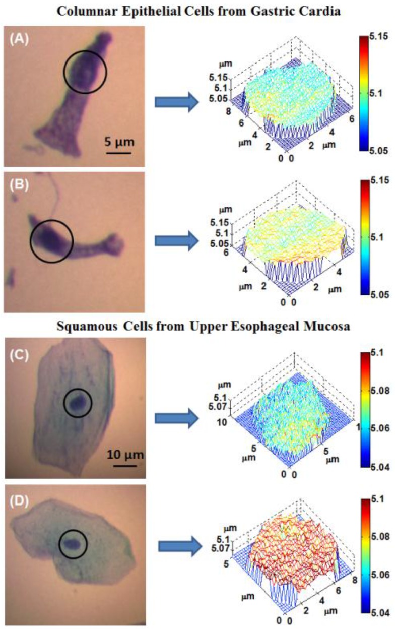
Patients with normal esophagus, gastroesophageal reflux, BE and EAC were eligible for enrollment. We performed endoscopic cytology brushings of the gastric cardia, ~1-2 cm below the gastroesophageal junction, and of the normal squamous esophageal mucosa at ~20 cm from the incisors and standard cytology slides were made using Thinprep method. Optical analysis was performed on the cell nuclei of cytologically normal-appearing epithelial cells.
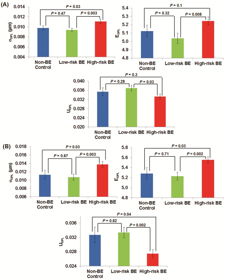
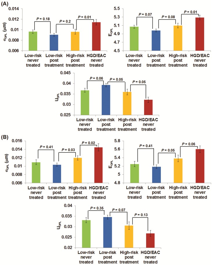
The study cohort consisted of 128 patients. The nuclear nano-architecture markers detected the presence of esophageal dysplasia and EAC with statistical significance. The field effect does not exhibit a spatial dependence. These markers reverted toward normal in response to endoscopic therapy.
Optical analysis of gastric cardia and upper squamous esophagus represents a potentially viable method to improve risk stratification and ease of surveillance of patients with Barrett's esophagus and to monitor the efficacy of ablative therapy.
巴雷特食管(BE)影响多达 1200 万美国人,使食管腺癌(EAC)的发展风险增加。EAC 通常是致命的,除非早期发现。鉴于其高患病率、高监测成本和大多数受影响个体的相对低风险,确定高危患者进行额外检查、定期监测或消融治疗至关重要。通过探查未受影响的食管黏膜来探索“场效应”,以预测 EAC 的风险,这可能是一种改进的监测和预防策略。在这项研究中,我们评估了来自正常鳞状食管和胃贲门的核纳米结构标记物检测食管异型增生和 EAC 的“场效应”及其对内镜治疗的反应的能力。
符合条件的患者为正常食管、胃食管反流、BE 和 EAC。我们对胃贲门(距食管胃交界处下方约 1-2cm)和距切牙约 20cm 的正常鳞状食管黏膜进行内镜细胞学刷检,并使用 Thinprep 方法制作标准细胞学载玻片。对细胞学表现正常的上皮细胞的细胞核进行光学分析。
研究队列包括 128 名患者。核纳米结构标记物检测到食管异型增生和 EAC 的存在具有统计学意义。场效应没有表现出空间依赖性。这些标记物在接受内镜治疗后恢复正常。
胃贲门和上食管鳞状上皮的光学分析可能是一种可行的方法,可以改善 Barrett 食管患者的风险分层和监测的便利性,并监测消融治疗的效果。