Department of Physics, University of Wisconsin-Milwaukee, 1900 E Kenwood Blvd, Milwaukee, WI 53211, USA.
Int J Mol Sci. 2013 Nov 19;14(11):22753-81. doi: 10.3390/ijms141122753.
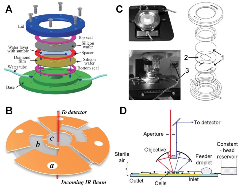
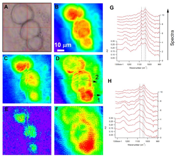
Infrared (IR) spectromicroscopy, or chemical imaging, is an evolving technique that is poised to make significant contributions in the fields of biology and medicine. Recent developments in sources, detectors, measurement techniques and speciman holders have now made diffraction-limited Fourier transform infrared (FTIR) imaging of cellular chemistry in living cells a reality. The availability of bright, broadband IR sources and large area, pixelated detectors facilitate live cell imaging, which requires rapid measurements using non-destructive probes. In this work, we review advances in the field of FTIR spectromicroscopy that have contributed to live-cell two and three-dimensional IR imaging, and discuss several key examples that highlight the utility of this technique for studying the structure and chemistry of living cells.
红外(IR)光谱显微镜,或化学成像,是一种不断发展的技术,有望在生物学和医学领域做出重大贡献。最近在光源、探测器、测量技术和标本支架方面的发展,使得对活细胞中细胞化学的衍射极限傅里叶变换红外(FTIR)成像成为现实。明亮、宽带的红外光源和大面积、像素化探测器的可用性促进了活细胞成像,这需要使用非破坏性探针进行快速测量。在这项工作中,我们回顾了 FTIR 光谱显微镜领域的进展,这些进展有助于对活细胞进行二维和三维 IR 成像,并讨论了几个关键示例,突出了该技术在研究活细胞结构和化学方面的应用。