Department of Biology, Institute for Marine Biosystems and Neurosciences, Shanghai Ocean University, Shanghai, 201306, China.
Cell Death Dis. 2013 Nov 21;4(11):e930. doi: 10.1038/cddis.2013.456.
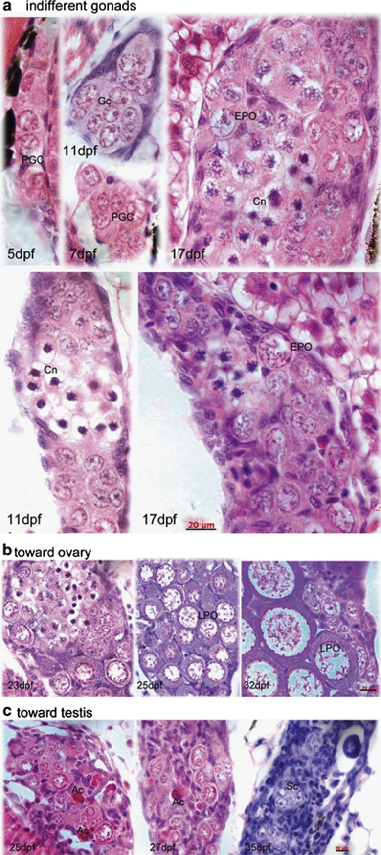
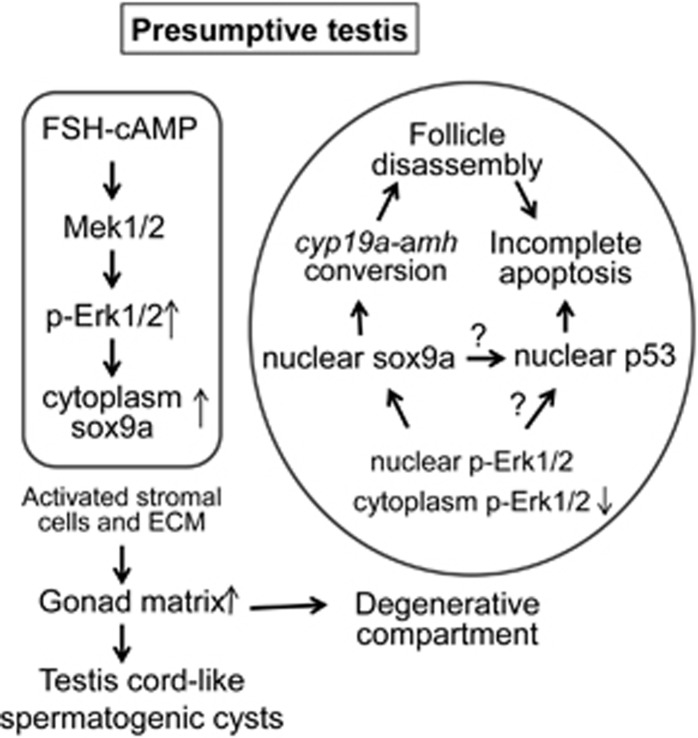
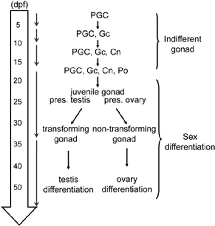
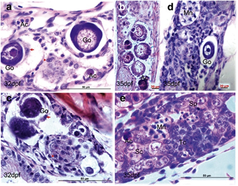
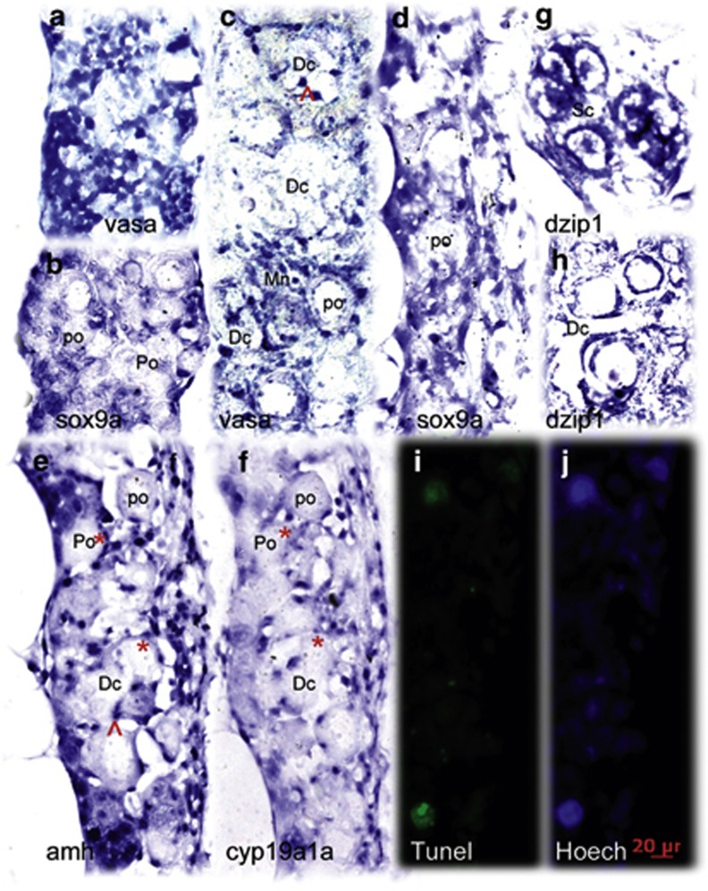
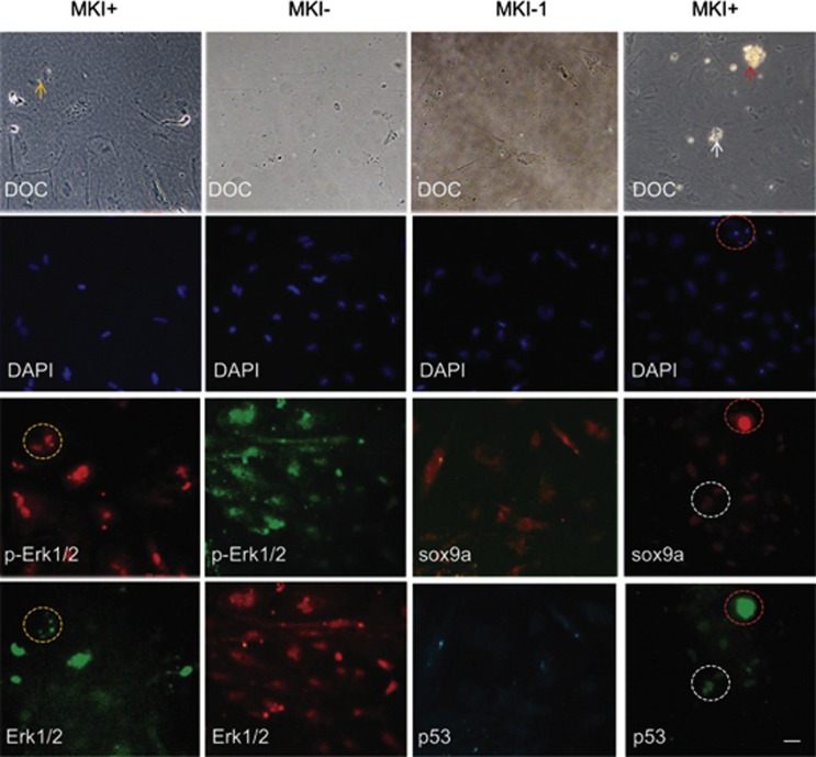
In almost all vertebrates, the downstream of the sox9 signaling axis is well conserved for testis differentiation. The upstream genes of this pathway vary from species to species during evolution. Yet, little is known about how these signaling cascades are regulated and what cellular processes are dominant in ovary-testis transformation in juvenile zebrafish. In this study, we find that the transforming gonads undergo activation of sox9a-expressing stromal cells with increased deposition of extracellular matrix and formation of degenerative compartments. This leads to follicle disassembly, oocyte degeneration, follicle cell-cyp19a1a-amh conversions, and, eventually, formation of the testis cord. In vitro primary culture of juvenile ovary tissue in gonadotropins increases cytoplasmic accumulation of sox9a and p-Erk1/2, and induces mesenchymal morphology. MAPK inhibitors (MKI), a mixture of PD98059 and U0216, eliminate the cytoplasmic distribution but do not eradicate nuclear localization of sox9a and p-Erk1/2. Nuclear p53 are relatively increased in MKI-treated cells that exhibit less spreading and reduced proliferation. Despite uniform nuclear condensation, only a fraction of cells displayed the apoptotic phenotype. These results suggest that high levels of cytoplasmic sox9a and p-Erk1/2 activity activate stromal cells and enhance the production of extracellular matrix required for testis cord formation, whereas deregulation and translocation of sox9a and p-Erk1/2 induce follicle disassembly and incomplete apoptosis associated with nuclear p53. Together with the established FSH/cAMP/MAPK/AMH pathway in mammalian granulosa and Sertoli cells, we demonstrated that the sox9 axis signaling that determines testis formation in mammals also induces zebrafish ovary-testis transition, and adds to its conserved role in sex reversal.
在几乎所有的脊椎动物中, Sox9 信号轴的下游对于睾丸分化是高度保守的。在进化过程中,该途径的上游基因在不同物种之间存在差异。然而,对于这些信号级联如何被调控以及在幼年斑马鱼的卵巢-睾丸转变中哪些细胞过程占主导地位,我们知之甚少。在这项研究中,我们发现,转化的生殖腺经历了 Sox9a 表达的基质细胞的激活,伴随着细胞外基质的沉积增加和退化隔室的形成。这导致卵泡解体、卵母细胞退化、卵泡细胞-cyp19a1a-amh 转化,最终形成睾丸索。在促性腺激素中对幼年卵巢组织进行体外原代培养会增加 Sox9a 和 p-Erk1/2 的细胞质积累,并诱导间充质形态。MAPK 抑制剂(MKI),即 PD98059 和 U0216 的混合物,消除了 Sox9a 和 p-Erk1/2 的细胞质分布,但不能消除核定位。在 MKI 处理的细胞中,p53 的核内相对增加,这些细胞表现出较少的扩散和降低的增殖。尽管核浓缩均匀,但只有一部分细胞表现出凋亡表型。这些结果表明,高水平的细胞质 Sox9a 和 p-Erk1/2 活性激活基质细胞,并增强形成睾丸索所需的细胞外基质的产生,而 Sox9a 和 p-Erk1/2 的失调和易位导致卵泡解体和与核内 p53 相关的不完全凋亡。与哺乳动物颗粒细胞和 Sertoli 细胞中已建立的 FSH/cAMP/MAPK/AMH 途径一起,我们证明了决定哺乳动物睾丸形成的 Sox9 轴信号也诱导了斑马鱼卵巢-睾丸转变,并增加了其在性别反转中的保守作用。