• 文献检索
  • 文档翻译
  • 深度研究
  • 学术资讯
  • Suppr Zotero 插件Zotero 插件
  • 邀请有礼
  • 套餐&价格
  • 历史记录
应用&插件
Suppr Zotero 插件Zotero 插件浏览器插件Mac 客户端Windows 客户端微信小程序
定价
高级版会员购买积分包购买API积分包
服务
文献检索文档翻译深度研究API 文档MCP 服务
关于我们
关于 Suppr公司介绍联系我们用户协议隐私条款
关注我们

Suppr 超能文献

核心技术专利:CN118964589B侵权必究
粤ICP备2023148730 号-1Suppr @ 2026

文献检索

告别复杂PubMed语法,用中文像聊天一样搜索,搜遍4000万医学文献。AI智能推荐,让科研检索更轻松。

立即免费搜索

文件翻译

保留排版,准确专业,支持PDF/Word/PPT等文件格式,支持 12+语言互译。

免费翻译文档

深度研究

AI帮你快速写综述,25分钟生成高质量综述,智能提取关键信息,辅助科研写作。

立即免费体验

葡萄膜炎患者的视网膜前膜:频域光学相干断层扫描的形态学和功能分析。

Epiretinal membranes in patients with uveitis: morphological and functional analysis with spectral domain optical coherence tomography.

机构信息

Department of Ophthalmology, Sapienza University of Rome, 15500161 Rome, Italy ; Servizio di Immunovirologia Oculare, Sapienza Università di Roma, Viale del Policlinico, 15500161 Rome, Italy.

出版信息

Biomed Res Int. 2013;2013:284821. doi: 10.1155/2013/284821. Epub 2013 Nov 5.

DOI:10.1155/2013/284821
PMID:24294602
原文链接:https://pmc.ncbi.nlm.nih.gov/articles/PMC3835473/
Abstract

PURPOSE

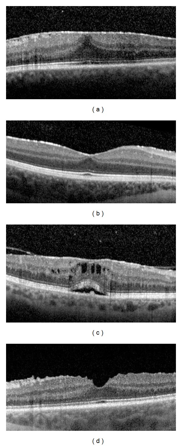
To correlate the uveitic epiretinal membrane (ERM) features using spectral-domain optical coherence tomography (SD-OCT) with visual acuity (VA).

METHODS

Forty-one eyes of 32 patients were included in this retrospective study. SD-OCT was performed in all patients and data were collected at the time of ERM diagnosis and at the final visit. Both best corrected visual acuity (BCVA) and ERM thickness were correlated with the morphological and clinical features.

RESULTS

Final BCVA was positively correlated with male sex (P = 0.0055) and the focal pattern of ERM attachment (P = 0.031) and negatively correlated with IS/OS photoreceptor junction disruption (P = 0.042). BVCA change showed a positive correlation with the age of ERM onset (P = 0.056) but a negative correlation with IS/OS photoreceptor disruption at the ERM diagnosis (P = 0.029) and the increase of central subfield thickness (CST) (P = 0.95). Final ERM thickness correlated with the duration of uveitis (P = 0.0023) and the duration of ERM (P = 1.15  e-05). During the follow-up, ERM thickening correlated with male sex (P = 0.042), posterior uveitis (P = 0.036), uveitis duration (P = 0.026), and broad attachment pattern (P = 0.052).

CONCLUSIONS

In the uveitic ERM, VA negatively correlates with IS/OS photoreceptor junction disruption and the increase of CST. ERM thickness is influenced by longer duration of both uveitis and ERM.

摘要

目的

通过频域光相干断层扫描(SD-OCT)与视力(VA)相关联,分析葡萄膜炎性视网膜内表面膜(ERM)的特征。

方法

本回顾性研究共纳入 32 例 41 只眼的患者。所有患者均行 SD-OCT 检查,并在 ERM 诊断时和最终就诊时采集数据。将最佳矫正视力(BCVA)和 ERM 厚度与形态学和临床特征相关联。

结果

最终 BCVA 与男性(P=0.0055)和 ERM 附着的局灶性模式(P=0.031)呈正相关,与 IS/OS 光感受器连接中断(P=0.042)呈负相关。BCVA 变化与 ERM 发病年龄呈正相关(P=0.056),但与 ERM 诊断时 IS/OS 光感受器破坏(P=0.029)和中央子场厚度(CST)增加(P=0.95)呈负相关。最终 ERM 厚度与葡萄膜炎持续时间(P=0.0023)和 ERM 持续时间(P=1.15×10⁻⁵)相关。在随访期间,ERM 增厚与男性(P=0.042)、后葡萄膜炎(P=0.036)、葡萄膜炎持续时间(P=0.026)和广泛附着模式(P=0.052)相关。

结论

在葡萄膜炎性 ERM 中,VA 与 IS/OS 光感受器连接中断和 CST 增加呈负相关。ERM 厚度受葡萄膜炎和 ERM 持续时间的影响。

https://cdn.ncbi.nlm.nih.gov/pmc/blobs/60ec/3835473/fb9d0ffd280b/BMRI2013-284821.003.jpg
https://cdn.ncbi.nlm.nih.gov/pmc/blobs/60ec/3835473/687348a4fa30/BMRI2013-284821.001.jpg
https://cdn.ncbi.nlm.nih.gov/pmc/blobs/60ec/3835473/ff64a333bb56/BMRI2013-284821.002.jpg
https://cdn.ncbi.nlm.nih.gov/pmc/blobs/60ec/3835473/fb9d0ffd280b/BMRI2013-284821.003.jpg
https://cdn.ncbi.nlm.nih.gov/pmc/blobs/60ec/3835473/687348a4fa30/BMRI2013-284821.001.jpg
https://cdn.ncbi.nlm.nih.gov/pmc/blobs/60ec/3835473/ff64a333bb56/BMRI2013-284821.002.jpg
https://cdn.ncbi.nlm.nih.gov/pmc/blobs/60ec/3835473/fb9d0ffd280b/BMRI2013-284821.003.jpg

相似文献

1
Epiretinal membranes in patients with uveitis: morphological and functional analysis with spectral domain optical coherence tomography.葡萄膜炎患者的视网膜前膜:频域光学相干断层扫描的形态学和功能分析。
Biomed Res Int. 2013;2013:284821. doi: 10.1155/2013/284821. Epub 2013 Nov 5.
2
Morphometric spectral-domain optical coherence tomography features of epiretinal membrane correlate with visual acuity in patients with uveitis.后发性葡萄膜炎患者的视网膜前膜形态计量谱域光相干断层扫描特征与视力相关。
Am J Ophthalmol. 2012 Jul;154(1):78-86.e1. doi: 10.1016/j.ajo.2012.01.032. Epub 2012 Apr 26.
3
Correlation between visual acuity changes and optical coherence tomography morphological findings in idiopathic epiretinal membranes.特发性视网膜前膜视力变化与光学相干断层扫描形态学结果的相关性
Graefes Arch Clin Exp Ophthalmol. 2016 Mar;254(3):437-44. doi: 10.1007/s00417-015-3069-0. Epub 2015 May 29.
4
Disruption of the photoreceptor inner segment/outer segment layer on spectral domain-optical coherence tomography is a predictor of poor visual acuity in patients with epiretinal membranes.在频域光相干断层扫描中,光感受器内节/外节层的破坏是预测视网膜前膜患者视力不良的指标。
Retina. 2010 May;30(5):713-8. doi: 10.1097/IAE.0b013e3181c596e3.
5
Associations between macular findings by optical coherence tomography and visual outcomes after epiretinal membrane removal.光学相干断层扫描黄斑区表现与视网膜前膜去除术后视觉预后的相关性。
Am J Ophthalmol. 2009 Mar;147(3):473-480.e3. doi: 10.1016/j.ajo.2008.09.020. Epub 2008 Dec 3.
6
Evaluation of inner segment/outer segment junctions in different types of epiretinal membranes.不同类型视网膜前膜内节/外节连接的评估
Arq Bras Oftalmol. 2016 Sep-Oct;79(5):319-322. doi: 10.5935/0004-2749.20160091.
7
Correlation of visual recovery with presence of photoreceptor inner/outer segment junction in optical coherence images after epiretinal membrane surgery.视网膜前膜手术后光学相干图像中光感受器内/外段连接的存在与视觉恢复的相关性。
Br J Ophthalmol. 2009 Feb;93(2):171-5. doi: 10.1136/bjo.2008.146381. Epub 2008 Oct 29.
8
Correlation between foveal cone outer segment tips line and visual recovery after epiretinal membrane surgery.黄斑区锥体细胞外节端线与视网膜前膜手术后视力恢复的相关性。
Invest Ophthalmol Vis Sci. 2013 Nov 5;54(12):7302-8. doi: 10.1167/iovs.13-12702.
9
Epiretinal membrane profile on spectral domain optical coherence tomography in patients with uveitis.葡萄膜炎患者频域光学相干断层扫描的视网膜内界膜特征。
Indian J Ophthalmol. 2019 Mar;67(3):376-381. doi: 10.4103/ijo.IJO_650_18.
10
Outer retinal morphology and visual function in patients with idiopathic epiretinal membrane.特发性眼内膜患者的视网膜外层形态和视觉功能。
JAMA Ophthalmol. 2013 Feb;131(2):172-7. doi: 10.1001/jamaophthalmol.2013.686.

引用本文的文献

1
Optical Coherence Tomography: Focus on the Pathology of Macula in Scleritis Patients.光学相干断层扫描:聚焦巩膜炎患者黄斑病变
J Clin Med. 2023 Jul 21;12(14):4825. doi: 10.3390/jcm12144825.
2
Correlation between Morphological Characteristics of Macular Edema and Visual Acuity in Young Patients with Idiopathic Intermediate Uveitis.特发性中间葡萄膜炎青年患者黄斑水肿形态特征与视力的相关性。
Medicina (Kaunas). 2023 Mar 8;59(3):529. doi: 10.3390/medicina59030529.
3
Functional and Anatomical Outcomes of Pars Plana Vitrectomy for Epiretinal Membrane in Patients with Uveitis.

本文引用的文献

1
Associations between metamorphopsia and foveal microstructure in patients with epiretinal membrane.视网膜前膜患者的形觉扭曲与中心凹微观结构之间的关系。
Invest Ophthalmol Vis Sci. 2012 Oct 3;53(11):6770-5. doi: 10.1167/iovs.12-9683.
2
Longitudinal morphometric analysis of epiretinal membrane in patients with uveitis.葡萄膜炎患者眼内视网膜膜的纵向形态计量分析。
Ocul Immunol Inflamm. 2013;21(1):2-7. doi: 10.3109/09273948.2012.714044. Epub 2012 Sep 4.
3
Morphometric spectral-domain optical coherence tomography features of epiretinal membrane correlate with visual acuity in patients with uveitis.
葡萄膜炎患者视网膜前膜的玻璃体切割术的功能和解剖学结果
Diagnostics (Basel). 2022 Dec 5;12(12):3044. doi: 10.3390/diagnostics12123044.
4
Spectral domain optical coherence tomography findings of patients with pars planitis and risk factors affecting visual acuity.特发性旁中心性脉络膜视网膜炎患者的光谱域光学相干断层扫描表现及影响视力的相关因素。
Int Ophthalmol. 2021 May;41(5):1753-1761. doi: 10.1007/s10792-021-01734-z. Epub 2021 Feb 9.
5
Epiretinal membrane: optical coherence tomography-based diagnosis and classification.视网膜前膜:基于光学相干断层扫描的诊断与分类
Clin Ophthalmol. 2016 Mar 29;10:527-34. doi: 10.2147/OPTH.S97722. eCollection 2016.
6
Correlation between visual acuity changes and optical coherence tomography morphological findings in idiopathic epiretinal membranes.特发性视网膜前膜视力变化与光学相干断层扫描形态学结果的相关性
Graefes Arch Clin Exp Ophthalmol. 2016 Mar;254(3):437-44. doi: 10.1007/s00417-015-3069-0. Epub 2015 May 29.
7
Photoreceptor inner segment ellipsoid band integrity on spectral domain optical coherence tomography.光谱域光学相干断层扫描上的光感受器内节椭圆体带完整性
Clin Ophthalmol. 2014 Dec 9;8:2507-22. doi: 10.2147/OPTH.S72132. eCollection 2014.
后发性葡萄膜炎患者的视网膜前膜形态计量谱域光相干断层扫描特征与视力相关。
Am J Ophthalmol. 2012 Jul;154(1):78-86.e1. doi: 10.1016/j.ajo.2012.01.032. Epub 2012 Apr 26.
4
Immunohistochemical study of epiretinal membranes in patients with uveitis.葡萄膜炎患者视网膜前膜的免疫组织化学研究
J Ophthalmic Inflamm Infect. 2012 Dec;2(4):243-8. doi: 10.1007/s12348-012-0074-x. Epub 2012 Apr 25.
5
Spectral-domain optical coherence tomography in uveitic macular edema: morphological features and prognostic factors.频域光相干断层扫描在葡萄膜炎性黄斑水肿中的应用:形态学特征和预后因素。
Ophthalmologica. 2012;228(1):13-8. doi: 10.1159/000337234. Epub 2012 Apr 14.
6
Morphologic and functional association of retinal layers beneath the epiretinal membrane with spectral-domain optical coherence tomography in eyes without photoreceptor abnormality.无感光细胞异常的眼内反膜下视网膜各层与频域光相干断层扫描的形态和功能关系。
Graefes Arch Clin Exp Ophthalmol. 2012 Apr;250(4):491-8. doi: 10.1007/s00417-011-1848-9. Epub 2011 Nov 16.
7
Role of the intravitreal growth factors in the pathogenesis of idiopathic epiretinal membrane.玻璃体内生长因子在特发性视网膜内表面膜发病机制中的作用。
Invest Ophthalmol Vis Sci. 2011 Jul 29;52(8):5786-9. doi: 10.1167/iovs.10-7116.
8
Preoperative inner segment/outer segment junction in spectral-domain optical coherence tomography as a prognostic factor in epiretinal membrane surgery.频域光学相干断层扫描术前内节/外节连接作为眼后膜手术的预后因素。
Retina. 2011 Jul-Aug;31(7):1366-72. doi: 10.1097/IAE.0b013e318203c156.
9
Inner segment/outer segment junction assessed by spectral-domain optical coherence tomography in patients with idiopathic epiretinal membrane.频域光相干断层扫描评估特发性视网膜前膜患者的内节/外节连接。
Am J Ophthalmol. 2010 Dec;150(6):834-9. doi: 10.1016/j.ajo.2010.06.006. Epub 2010 Aug 17.
10
Spectral-domain optical coherence tomography use in macular diseases: a review.谱域光学相干断层扫描在黄斑疾病中的应用:综述。
Ophthalmologica. 2010;224(6):333-40. doi: 10.1159/000313814. Epub 2010 May 4.