Ni Jing-qin, Ouyang Qiufang, Lin Ling, Huang Ziyang, Lu Huixia, Chen Xiaoqing, Lin Huili, Wang Zhenhua, Xu Dongming, Zhang Yun
Cardiovascular Department, Second Affiliated Hospital and Second Clinical Medical College, Fujian Medical University, Quanzhou, Fujian 362000, China.
Clin Dev Immunol. 2013;2013:476856. doi: 10.1155/2013/476856. Epub 2013 Sep 15.
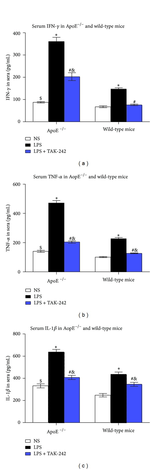
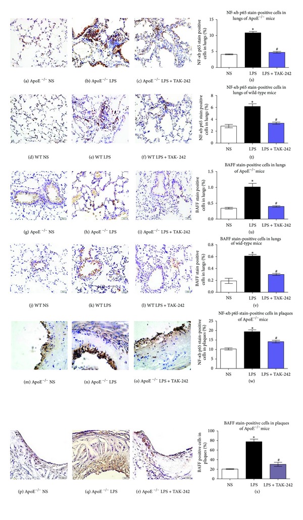
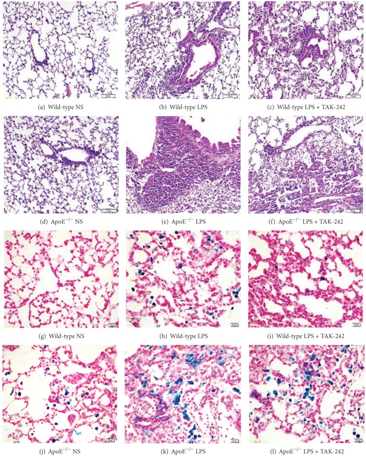
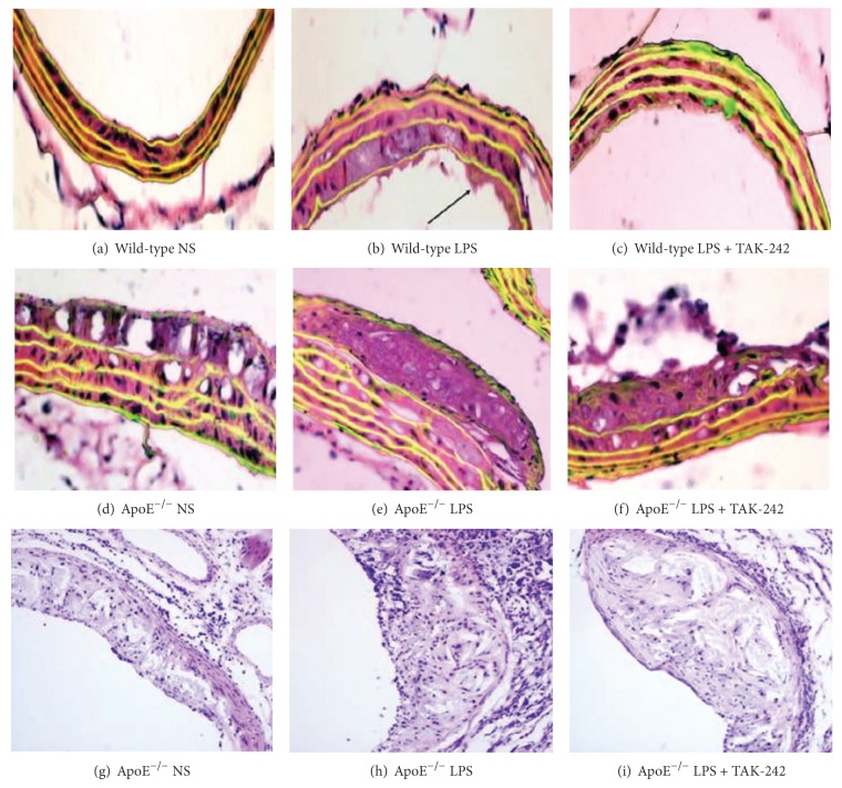
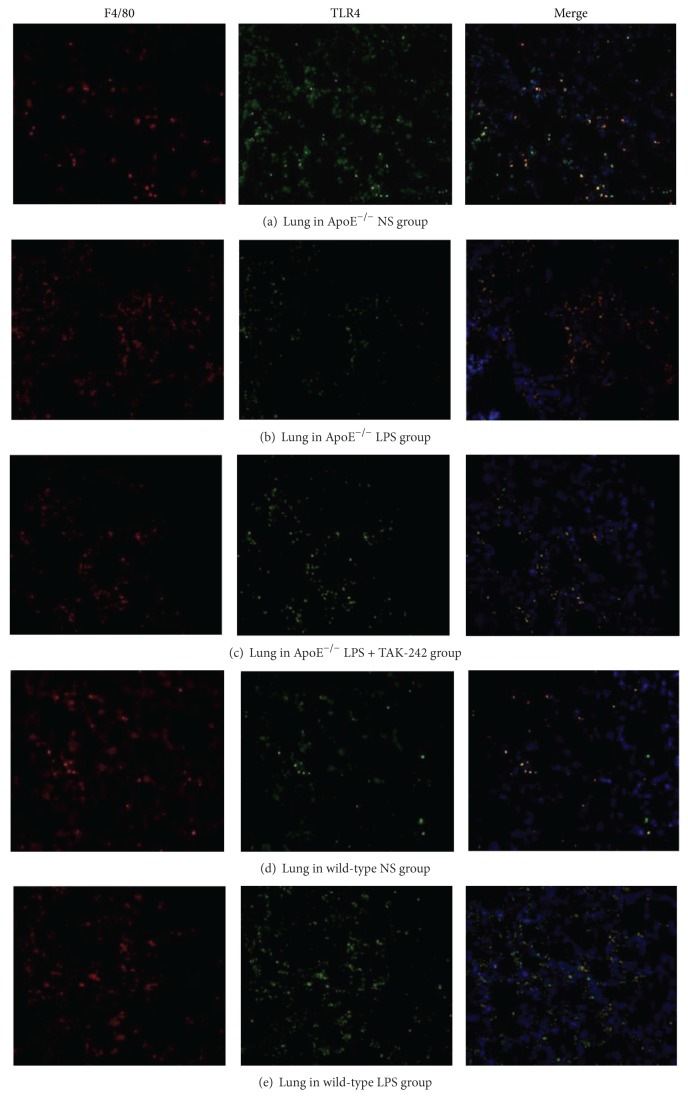
To investigate the pathologic mechanisms of toll-like receptor 4 (TLR4) in lung injury and atherosclerosis, ApoE⁻/⁻ or wild-type mice were intraperitoneally administered saline, lipopolysaccharides (LPS), or LPS plus TAK-242 (TLR4 inhibitor), respectively, twice a week for 4 weeks. Serum autoantibody of antinuclear antibody (ANA), anti-double-stranded DNA (anti-dsDNA), and cytokines of interferon-gamma (IFN-γ), tumor necrosis factor (TNF-α ), and interleukin-1 (IL-1β) were assessed by ELISA. Hematoxylin and eosin (HE) and Perl's stains for lung pathomorphology as well as HE staining for atherosclerosis were employed. TLR4 in macrophages was detected by double immunofluorescent staining. While protein expressions of TLR4, nuclear factor-kappa B p65 (NF-κB p65), and B cell activating factor belonging to the TNF family (BAFF) were examined by immunohistochemistry. We found that serum autoantibody (ANA and anti-dsDNA), cytokines (IFN-γ, TNF-α, IL-1β), lung inflammation, and intima-media thickness in brachiocephalic artery were obviously increased after LPS challenge in both genotypes, but to a lesser extent in wild-type strains. And those alterations were alleviated by coadministration of LPS and TAK-242. Mechanistically, upregulation of TLR4, NF-κb, and BAFF was involved. We concluded that TLR4/NF-κb/BAFF in macrophages might be a possible common autoimmune pathway that caused lung injury and atherosclerosis. TLR4 signal will be a therapeutic target in atherosclerosis and immune-mediated lung injury.
为研究Toll样受体4(TLR4)在肺损伤和动脉粥样硬化中的病理机制,分别给载脂蛋白E基因敲除(ApoE⁻/⁻)小鼠或野生型小鼠腹腔注射生理盐水、脂多糖(LPS)或LPS加TAK-242(TLR4抑制剂),每周两次,共4周。采用酶联免疫吸附测定(ELISA)法检测血清抗核抗体(ANA)、抗双链DNA抗体(抗dsDNA)自身抗体以及干扰素-γ(IFN-γ)、肿瘤坏死因子(TNF-α)和白细胞介素-1(IL-1β)细胞因子。采用苏木精-伊红(HE)染色及Perl染色观察肺组织病理形态,采用HE染色观察动脉粥样硬化情况。通过双重免疫荧光染色检测巨噬细胞中的TLR4。采用免疫组织化学法检测TLR4、核因子-κB p65(NF-κB p65)和肿瘤坏死因子家族成员B细胞活化因子(BAFF)的蛋白表达。我们发现,两种基因型小鼠在LPS刺激后,血清自身抗体(ANA和抗dsDNA)、细胞因子(IFN-γ、TNF-α、IL-1β)、肺部炎症以及头臂动脉内膜中层厚度均明显增加,但野生型小鼠增加程度较小。LPS与TAK-242联合给药可减轻这些改变。机制上,涉及TLR4、NF-κB和BAFF的上调。我们得出结论,巨噬细胞中的TLR4/NF-κB/BAFF可能是导致肺损伤和动脉粥样硬化的一条可能的共同自身免疫途径。TLR4信号将成为动脉粥样硬化和免疫介导性肺损伤的治疗靶点。