Department of Pediatrics I, University Children's Hospital, University of Heidelberg, Heidelberg, Germany ;
PLoS One. 2013 Dec 4;8(12):e82137. doi: 10.1371/journal.pone.0082137. eCollection 2013.
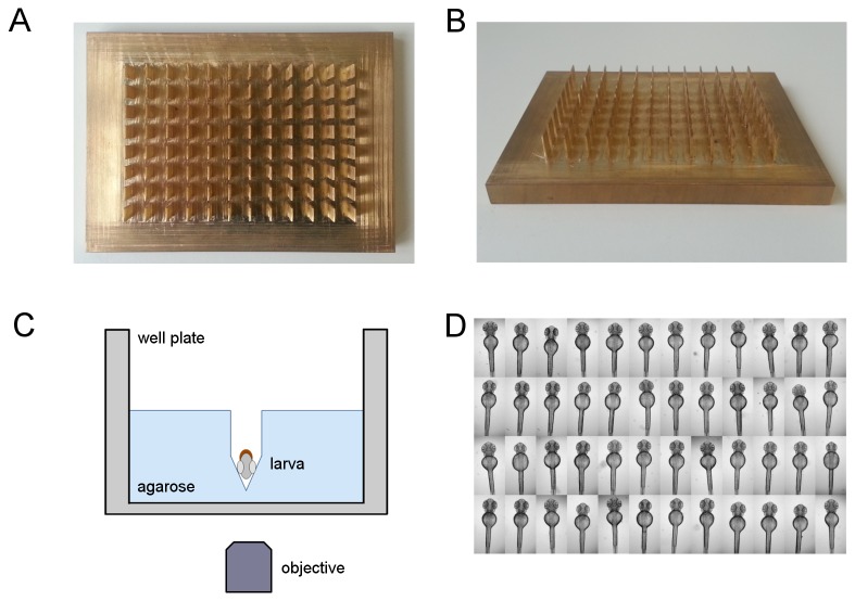
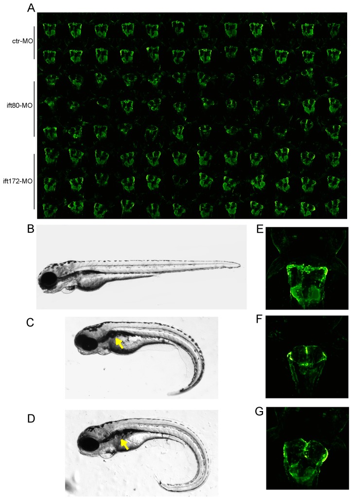
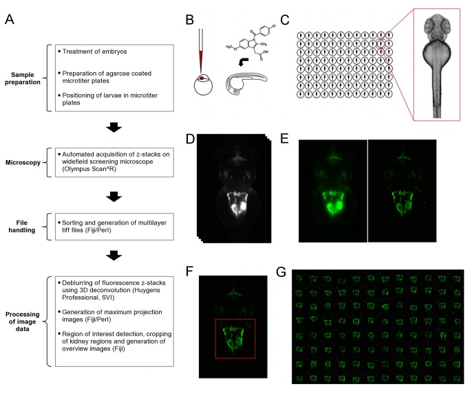
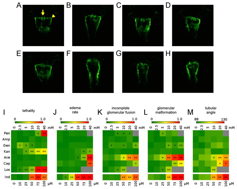
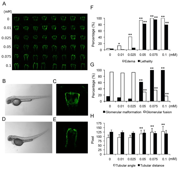
The analysis of kidney malformation caused by environmental influences during nephrogenesis or by hereditary nephropathies requires animal models allowing the in vivo observation of developmental processes. The zebrafish has emerged as a useful model system for the analysis of vertebrate organ development and function, and it is suitable for the identification of organotoxic or disease-modulating compounds on a larger scale. However, to fully exploit its potential in high content screening applications, dedicated protocols are required allowing the consistent visualization of inner organs such as the embryonic kidney. To this end, we developed a high content screening compatible pipeline for the automated imaging of standardized views of the developing pronephros in zebrafish larvae. Using a custom designed tool, cavities were generated in agarose coated microtiter plates allowing for accurate positioning and orientation of zebrafish larvae. This enabled the subsequent automated acquisition of stable and consistent dorsal views of pronephric kidneys. The established pipeline was applied in a pilot screen for the analysis of the impact of potentially nephrotoxic drugs on zebrafish pronephros development in the Tg(wt1b:EGFP) transgenic line in which the developing pronephros is highlighted by GFP expression. The consistent image data that was acquired allowed for quantification of gross morphological pronephric phenotypes, revealing concentration dependent effects of several compounds on nephrogenesis. In addition, applicability of the imaging pipeline was further confirmed in a morpholino based model for cilia-associated human genetic disorders associated with different intraflagellar transport genes. The developed tools and pipeline can be used to study various aspects in zebrafish kidney research, and can be readily adapted for the analysis of other organ systems.
分析肾畸形是由肾发生期间的环境影响引起的,还是由遗传性肾病引起的,需要动物模型来允许对发育过程进行体内观察。斑马鱼已成为分析脊椎动物器官发育和功能的有用模型系统,并且适合大规模鉴定器官毒性或疾病调节化合物。然而,要充分利用其在高通量筛选应用中的潜力,需要专门的协议来允许一致地可视化内部器官,如胚胎肾脏。为此,我们开发了一种高通量筛选兼容的流水线,用于自动成像斑马鱼幼虫中发育的前肾的标准化视图。使用定制设计的工具,在琼脂糖涂层的微孔板中产生腔,从而可以准确定位和定向斑马鱼幼虫。这使得随后可以自动获取稳定且一致的前肾背视图。所建立的流水线应用于初步筛选中,以分析潜在肾毒性药物对 Tg(wt1b:EGFP)转基因系中斑马鱼前肾发育的影响,其中 GFP 表达突出显示了发育中的前肾。所获得的一致图像数据允许对大体形态前肾表型进行定量,揭示了几种化合物对肾发生的浓度依赖性影响。此外,该成像流水线在基于 morpholino 的与人纤毛相关的遗传疾病模型中的适用性也得到了进一步证实,该模型与不同的鞭毛内运输基因有关。开发的工具和流水线可用于研究斑马鱼肾脏研究的各个方面,并且可以很容易地适应其他器官系统的分析。