• 文献检索
  • 文档翻译
  • 深度研究
  • 学术资讯
  • Suppr Zotero 插件Zotero 插件
  • 邀请有礼
  • 套餐&价格
  • 历史记录
应用&插件
Suppr Zotero 插件Zotero 插件浏览器插件Mac 客户端Windows 客户端微信小程序
定价
高级版会员购买积分包购买API积分包
服务
文献检索文档翻译深度研究API 文档MCP 服务
关于我们
关于 Suppr公司介绍联系我们用户协议隐私条款
关注我们

Suppr 超能文献

核心技术专利:CN118964589B侵权必究
粤ICP备2023148730 号-1Suppr @ 2026

文献检索

告别复杂PubMed语法,用中文像聊天一样搜索,搜遍4000万医学文献。AI智能推荐,让科研检索更轻松。

立即免费搜索

文件翻译

保留排版,准确专业,支持PDF/Word/PPT等文件格式,支持 12+语言互译。

免费翻译文档

深度研究

AI帮你快速写综述,25分钟生成高质量综述,智能提取关键信息,辅助科研写作。

立即免费体验

一种评估掌骨头、掌骨干和桡骨远端骨质量的定制方案:高分辨率外周定量 CT 精准研究。

A customized protocol to assess bone quality in the metacarpal head, metacarpal shaft and distal radius: a high resolution peripheral quantitative computed tomography precision study.

机构信息

Department of Physical Therapy, Faculty of Medicine, University of British Columbia (UBC), Vancouver, BC, Canada.

出版信息

BMC Musculoskelet Disord. 2013 Dec 24;14:367. doi: 10.1186/1471-2474-14-367.

DOI:10.1186/1471-2474-14-367
PMID:24364867
原文链接:https://pmc.ncbi.nlm.nih.gov/articles/PMC3877978/
Abstract

BACKGROUND

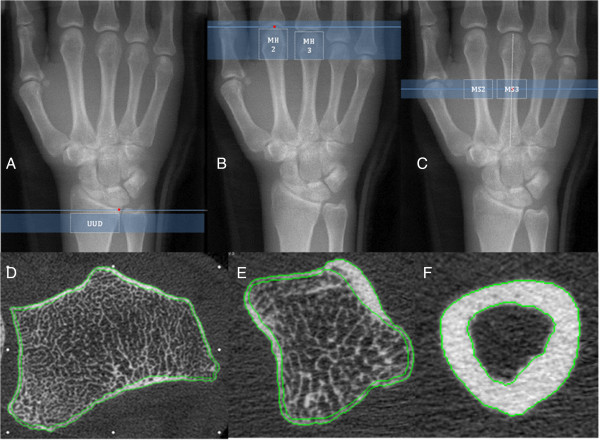
High Resolution-Peripheral Quantitative Computed Tomography (HR-pQCT) is an emerging technology for evaluation of bone quality in Rheumatoid Arthritis (RA). However, there are limitations with standard HR-pQCT imaging protocols for examination of regions of bone commonly affected in RA. We developed a customized protocol for evaluation of volumetric bone mineral density (vBMD) and microstructure at the metacarpal head (MH), metacarpal shaft (MS) and ultra-ultra-distal (UUD) radius; three sites commonly affected in RA. The purpose was to evaluate short-term measurement precision for bone density and microstructure at these sites.

METHODS

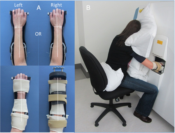
12 non-RA participants, individuals likely to have no pre-existing bone damage, consented to participate [8 females, aged 23 to 71 y [median (IQR): 44 (28) y]. The custom protocol includes more comfortable/stable positioning and adapted cortical segmentation and direct transformation analysis methods. Dominant arm MH, MS and UUD radius scans were completed on day one; repeated twice (with repositioning) three to seven days later. Short-term precision for repeated measures was explored using intraclass correlational coefficient (ICC), mean coefficient of variation (CV%), root mean square coefficient of variation (RMSCV%) and least significant change (LSC%95).

RESULTS

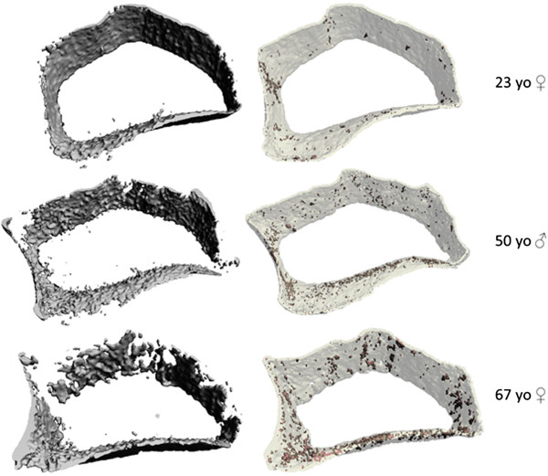
Bone density and microstructure precision was excellent: ICCs varied from 0.88 (MH2 trabecular number) to .99 (MS3 polar moment of inertia); CV% varied from < 1 (MS2 vBMD) to 6 (MS3 marrow space diameter); RMSCV% varied from < 1 (MH2 full bone vBMD) to 7 (MS3 marrow space diameter); and LSC%95 varied from 2 (MS2 full bone vBMD to 21 (MS3 marrow space diameter). Cortical porosity measures were the exception; RMSCV% varying from 19 (MS3) to 42 (UUD). No scans were stopped for discomfort. 5% (5/104) were repeated due to motion during imaging. 8% (8/104) of final images had motion artifact graded > 3 on 5 point scale.

CONCLUSION

In our facility, this custom protocol extends the potential for in vivo HR-pQCT imaging to assess, with high precision, regional differences in bone quality at three sites commonly affected in RA. Our methods are easy to adopt and we recommend other users of HR-pQCT consider this protocol for further evaluations of its precision and feasibility in their imaging facilities.

摘要

背景

高分辨率外周定量计算机断层扫描(HR-pQCT)是一种新兴的技术,可用于评估类风湿关节炎(RA)中的骨质量。然而,对于检查 RA 常见受累部位的骨,标准的 HR-pQCT 成像方案存在局限性。我们开发了一种定制方案,用于评估掌骨头部(MH)、掌骨干(MS)和超远端(UUD)桡骨的容积骨矿物质密度(vBMD)和微观结构;这三个部位是 RA 常见的受累部位。其目的是评估这些部位骨密度和微观结构的短期测量精度。

方法

12 名非 RA 参与者(8 名女性,年龄 23 岁至 71 岁[中位数(IQR):44(28)岁])同意参加研究。该定制方案包括更舒适/稳定的定位和适应的皮质分割和直接转换分析方法。在第一天完成优势手臂 MH、MS 和 UUD 桡骨扫描;在三到七天后重复两次(重新定位)。使用组内相关系数(ICC)、平均变异系数(CV%)、均方根变异系数(RMSCV%)和最小显著变化(LSC%95)来探讨重复性测量的短期精度。

结果

骨密度和微观结构精度非常好:ICC 从 0.88(MH2 骨小梁数量)到 0.99(MS3 极惯性矩);CV%从 <1(MS2 vBMD)到 6(MS3 骨髓腔直径);RMSCV%从 <1(MH2 全骨 vBMD)到 7(MS3 骨髓腔直径);LSC%95 从 2(MS2 全骨 vBMD)到 21(MS3 骨髓腔直径)。皮质孔隙度测量是个例外;RMSCV%从 19(MS3)到 42(UUD)。没有因不适而停止扫描。5%(5/104)因成像过程中的运动而重复。8%(8/104)的最终图像在 5 分制的运动伪影评分为 >3。

结论

在我们的机构中,该定制方案扩展了 HR-pQCT 成像的应用潜力,可高精度评估 RA 常见受累的三个部位的骨质量区域差异。我们的方法易于采用,我们建议 HR-pQCT 的其他使用者考虑该方案,以进一步评估其在其成像设施中的精度和可行性。

https://cdn.ncbi.nlm.nih.gov/pmc/blobs/8ea3/3877978/483a3712f181/1471-2474-14-367-3.jpg
https://cdn.ncbi.nlm.nih.gov/pmc/blobs/8ea3/3877978/7ef7963f7bff/1471-2474-14-367-1.jpg
https://cdn.ncbi.nlm.nih.gov/pmc/blobs/8ea3/3877978/476dcec956b0/1471-2474-14-367-2.jpg
https://cdn.ncbi.nlm.nih.gov/pmc/blobs/8ea3/3877978/483a3712f181/1471-2474-14-367-3.jpg
https://cdn.ncbi.nlm.nih.gov/pmc/blobs/8ea3/3877978/7ef7963f7bff/1471-2474-14-367-1.jpg
https://cdn.ncbi.nlm.nih.gov/pmc/blobs/8ea3/3877978/476dcec956b0/1471-2474-14-367-2.jpg
https://cdn.ncbi.nlm.nih.gov/pmc/blobs/8ea3/3877978/483a3712f181/1471-2474-14-367-3.jpg

相似文献

1
A customized protocol to assess bone quality in the metacarpal head, metacarpal shaft and distal radius: a high resolution peripheral quantitative computed tomography precision study.一种评估掌骨头、掌骨干和桡骨远端骨质量的定制方案:高分辨率外周定量 CT 精准研究。
BMC Musculoskelet Disord. 2013 Dec 24;14:367. doi: 10.1186/1471-2474-14-367.
2
Micro-structural bone changes in early rheumatoid arthritis persist over 1-year despite use of disease modifying anti-rheumatic drug therapy.尽管使用了改善病情抗风湿药物治疗,但早期类风湿关节炎患者的骨微结构变化仍会持续1年以上。
BMC Musculoskelet Disord. 2017 Dec 11;18(1):521. doi: 10.1186/s12891-017-1888-3.
3
Quantitative characterization of metacarpal and radial bone in rheumatoid arthritis using high resolution- peripheral quantitative computed tomography.使用高分辨率外周定量计算机断层扫描对类风湿关节炎患者的掌骨和桡骨进行定量表征。
Int J Rheum Dis. 2017 Mar;20(3):353-362. doi: 10.1111/1756-185X.12558. Epub 2015 Apr 10.
4
Reduced trabecular bone mineral density and cortical thickness accompanied by increased outer bone circumference in metacarpal bone of rheumatoid arthritis patients: a cross-sectional study.类风湿关节炎患者掌骨的骨小梁骨密度和皮质厚度降低,而外骨周长增加:一项横断面研究。
Arthritis Res Ther. 2010;12(3):R119. doi: 10.1186/ar3056. Epub 2010 Jun 21.
5
Alterations of bone density, microstructure, and strength of the distal radius in male patients with rheumatoid arthritis: a case-control study with HR-pQCT.类风湿关节炎男性患者桡骨远端骨密度、微观结构及强度的改变:一项采用高分辨率外周定量CT的病例对照研究
J Bone Miner Res. 2014 Sep;29(9):2118-29. doi: 10.1002/jbmr.2221.
6
Bone density and microarchitecture: relationship between hand, peripheral, and axial skeletal sites assessed by HR-pQCT and DXA in rheumatoid arthritis.骨密度和微结构:类风湿关节炎患者通过高分辨率外周定量 CT(HR-pQCT)和双能 X 线吸收法(DXA)评估手部、外周和轴向骨骼部位之间的关系。
Calcif Tissue Int. 2012 Nov;91(5):343-55. doi: 10.1007/s00223-012-9644-z. Epub 2012 Sep 4.
7
Assessment of hand bone loss in rheumatoid arthritis by high-resolution peripheral quantitative CT.采用高分辨率外周定量 CT 评估类风湿关节炎患者手部骨丢失。
Ann Rheum Dis. 2010 Sep;69(9):1671-6. doi: 10.1136/ard.2009.114512. Epub 2010 Jun 4.
8
Structure and strength of the distal radius in female patients with rheumatoid arthritis: a case-control study.类风湿关节炎女性患者桡骨远端的结构和强度:病例对照研究。
J Bone Miner Res. 2013 Apr;28(4):794-806. doi: 10.1002/jbmr.1793.
9
Reproducible metacarpal joint space width measurements using 3D analysis of images acquired with high-resolution peripheral quantitative computed tomography.使用高分辨率外周定量计算机断层扫描获取的图像进行 3D 分析,可实现掌指关节间隙宽度的可重现测量。
Med Eng Phys. 2013 Oct;35(10):1540-4. doi: 10.1016/j.medengphy.2013.04.003. Epub 2013 May 14.
10
Age- and Sex-Dependent Changes of Intra-articular Cortical and Trabecular Bone Structure and the Effects of Rheumatoid Arthritis.关节内皮质骨和松质骨结构的年龄和性别依赖性变化及其对类风湿关节炎的影响。
J Bone Miner Res. 2017 Apr;32(4):722-730. doi: 10.1002/jbmr.3025. Epub 2016 Nov 18.

引用本文的文献

1
Peripheral quantitative computed tomography in the assessment of bone mineral density in anti-TNF-treated rheumatoid arthritis and ankylosing spondylitis patients.外周定量计算机断层扫描在评价抗 TNF 治疗的类风湿关节炎和强直性脊柱炎患者骨密度中的应用。
BMC Musculoskelet Disord. 2021 Sep 23;22(1):817. doi: 10.1186/s12891-021-04708-5.
2
3-D microarchitectural properties and rod- and plate-like trabecular morphometric properties of femur head cancellous bones in patients with rheumatoid arthritis, osteoarthritis, and osteoporosis.类风湿性关节炎、骨关节炎和骨质疏松症患者股骨头松质骨的三维微观结构特性以及杆状和板状小梁形态计量学特性。
J Orthop Translat. 2021 Apr 20;28:159-168. doi: 10.1016/j.jot.2021.02.002. eCollection 2021 May.
3

本文引用的文献

1
Rheumatoid arthritis of the hand: a five-year longitudinal analysis of clinical and radiographic findings.手部类风湿关节炎:五年临床和影像学研究。
Mod Rheumatol. 2014 Jan;24(1):69-77. doi: 10.3109/14397595.2013.854054.
2
Endocortical bone loss in osteoporosis: the role of bone surface availability.骨质疏松症中的内皮质骨丢失:骨表面可用性的作用。
Int J Numer Method Biomed Eng. 2013 Dec;29(12):1307-22. doi: 10.1002/cnm.2567. Epub 2013 Jul 1.
3
Can bone loss in rheumatoid arthritis be prevented?类风湿关节炎导致的骨质流失能否预防?
Pseudoerosions of Hands and Feet in Rheumatoid Arthritis: Anatomic Concepts and Redefinition.
类风湿关节炎中手足的假性糜烂:解剖学概念与重新定义
J Clin Med. 2019 Dec 9;8(12):2174. doi: 10.3390/jcm8122174.
4
Temporomandibular Disorders and Oral Features in Early Rheumatoid Arthritis Patients: An Observational Study.颞下颌关节紊乱与早期类风湿关节炎患者的口腔特征:一项观察性研究。
Int J Med Sci. 2019 Jan 1;16(2):253-263. doi: 10.7150/ijms.28361. eCollection 2019.
5
An automated algorithm for the detection of cortical interruptions and its underlying loss of trabecular bone; a reproducibility study.一种用于检测皮质中断及其潜在小梁骨丢失的自动化算法;一项重复性研究。
BMC Med Imaging. 2018 May 15;18(1):13. doi: 10.1186/s12880-018-0255-7.
6
Micro-structural bone changes in early rheumatoid arthritis persist over 1-year despite use of disease modifying anti-rheumatic drug therapy.尽管使用了改善病情抗风湿药物治疗,但早期类风湿关节炎患者的骨微结构变化仍会持续1年以上。
BMC Musculoskelet Disord. 2017 Dec 11;18(1):521. doi: 10.1186/s12891-017-1888-3.
7
Vascular channels in metacarpophalangeal joints: a comparative histologic and high-resolution imaging study.掌指关节中的血管通道:一项比较组织学和高分辨率成像研究。
Sci Rep. 2017 Aug 21;7(1):8966. doi: 10.1038/s41598-017-09363-2.
8
Diagnostic value of high-frequency color Doppler ultrasonography examination in combination with anti-cyclic citrullinated peptide antibody testing in rheumatoid arthritis patients.高频彩色多普勒超声检查联合抗环瓜氨酸肽抗体检测在类风湿关节炎患者中的诊断价值
Exp Ther Med. 2017 Mar;13(3):905-908. doi: 10.3892/etm.2017.4056. Epub 2017 Jan 17.
9
Development of three-dimensional prints of arthritic joints for supporting patients' awareness to structural damage.开发关节炎关节的三维打印模型以提高患者对结构损伤的认知。
Arthritis Res Ther. 2017 Feb 10;19(1):34. doi: 10.1186/s13075-017-1234-z.
10
Visual detection of cortical breaks in hand joints: reliability and validity of high-resolution peripheral quantitative CT compared to microCT.手部关节皮质骨断裂的视觉检测:高分辨率外周定量CT与显微CT相比的可靠性和有效性
BMC Musculoskelet Disord. 2016 Jul 11;17:271. doi: 10.1186/s12891-016-1148-y.
Osteoporos Int. 2013 Oct;24(10):2541-53. doi: 10.1007/s00198-013-2334-5. Epub 2013 Jun 18.
4
Catabolic and anabolic periarticular bone changes in patients with rheumatoid arthritis: a computed tomography study on the role of age, disease duration and bone markers.类风湿关节炎患者关节周围骨的分解代谢和合成代谢变化:一项关于年龄、病程和骨标志物作用的计算机断层扫描研究
Arthritis Res Ther. 2013;15(3):R62. doi: 10.1186/ar4235.
5
Reproducible metacarpal joint space width measurements using 3D analysis of images acquired with high-resolution peripheral quantitative computed tomography.使用高分辨率外周定量计算机断层扫描获取的图像进行 3D 分析,可实现掌指关节间隙宽度的可重现测量。
Med Eng Phys. 2013 Oct;35(10):1540-4. doi: 10.1016/j.medengphy.2013.04.003. Epub 2013 May 14.
6
Fracture history of healthy premenopausal women is associated with a reduction of cortical microstructural components at the distal radius.健康绝经前女性的骨折史与桡骨远端皮质微结构成分减少有关。
Bone. 2013 Aug;55(2):377-83. doi: 10.1016/j.bone.2013.04.025. Epub 2013 May 6.
7
Architecture of cortical bone determines in part its remodelling and structural decay.皮质骨的结构决定了其重塑和结构衰变的部分程度。
Bone. 2013 Aug;55(2):353-8. doi: 10.1016/j.bone.2013.04.020. Epub 2013 May 1.
8
Age-related changes in trabecular and cortical bone microstructure.与年龄相关的小梁骨和皮质骨微观结构的变化。
Int J Endocrinol. 2013;2013:213234. doi: 10.1155/2013/213234. Epub 2013 Mar 18.
9
Sites, frequencies, and causes of self-reported fractures in 9,720 rheumatoid arthritis patients: a large prospective observational cohort study in Japan.9720 例类风湿关节炎患者自述骨折的部位、频率和原因:日本一项大型前瞻性观察队列研究。
Arch Osteoporos. 2013;8:130. doi: 10.1007/s11657-013-0130-7. Epub 2013 Mar 23.
10
High-resolution peripheral quantitative computed tomography for the assessment of bone strength and structure: a review by the Canadian Bone Strength Working Group.高分辨率外周定量计算机断层扫描评估骨强度和结构:加拿大骨强度工作组综述。
Curr Osteoporos Rep. 2013 Jun;11(2):136-46. doi: 10.1007/s11914-013-0140-9.