Cardemil Carina, Elgali Ibrahim, Xia Wei, Emanuelsson Lena, Norlindh Birgitta, Omar Omar, Thomsen Peter
Department of Biomaterials, Institute of Clinical Sciences, Sahlgrenska Academy at University of Gothenburg, Gothenburg, Sweden ; Department of Oral and Maxillofacial Surgery, Örebro University Hospital, Örebro, Sweden ; BIOMATCELL VINN Excellence Center of Biomaterials and Cell Therapy, Gothenburg, Sweden.
Department of Biomaterials, Institute of Clinical Sciences, Sahlgrenska Academy at University of Gothenburg, Gothenburg, Sweden ; BIOMATCELL VINN Excellence Center of Biomaterials and Cell Therapy, Gothenburg, Sweden.
PLoS One. 2013 Dec 23;8(12):e84932. doi: 10.1371/journal.pone.0084932. eCollection 2013.
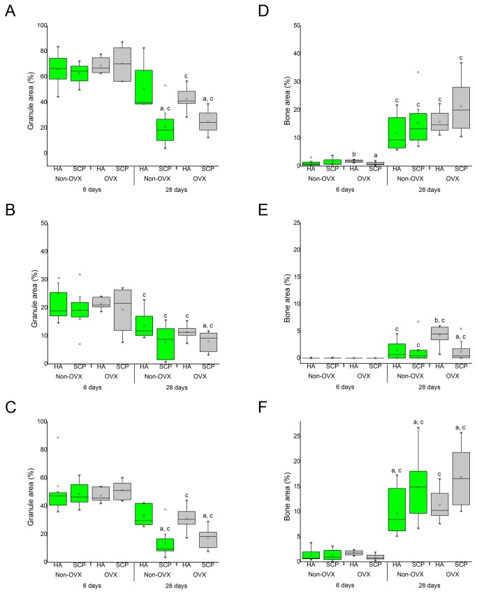
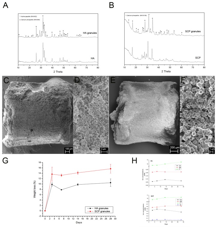
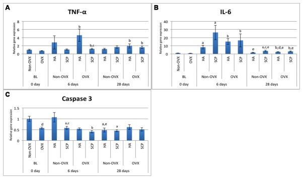
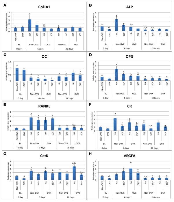
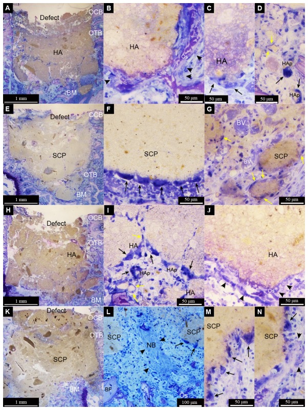
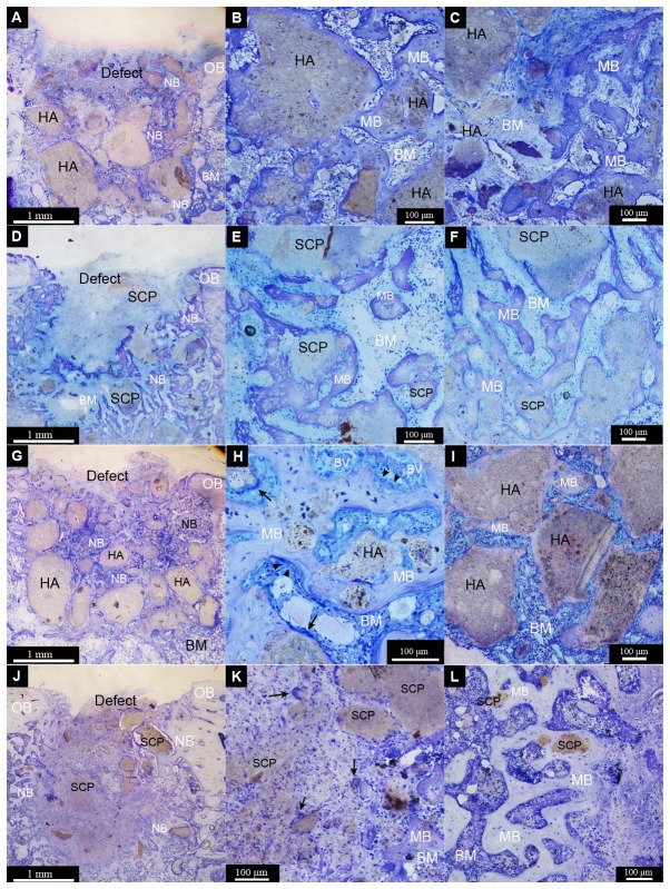
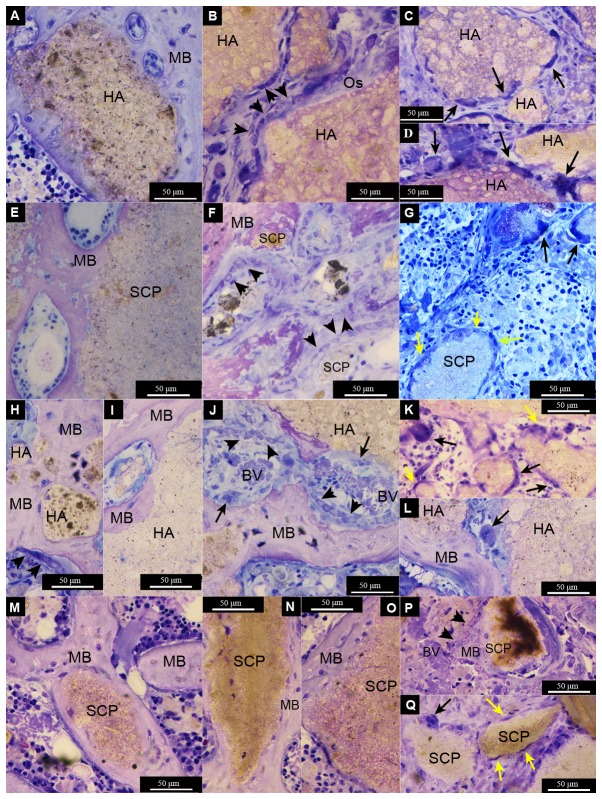
The healing of bone defects may be hindered by systemic conditions such as osteoporosis. Calcium phosphates, with or without ion substitutions, may provide advantages for bone augmentation. However, the mechanism of bone formation with these materials is unclear. The aim of this study was to evaluate the healing process in bone defects implanted with hydroxyapatite (HA) or strontium-doped calcium phosphate (SCP) granules, in non-ovariectomised (non-OVX) and ovariectomised (OVX) rats. After 0 (baseline), six and 28d, bone samples were harvested for gene expression analysis, histology and histomorphometry. Tumour necrosis factor-α (TNF-α), at six days, was higher in the HA, in non-OVX and OVX, whereas interleukin-6 (IL-6), at six and 28d, was higher in SCP, but only in non-OVX. Both materials produced a similar expression of the receptor activator of nuclear factor kappa-B ligand (RANKL). Higher expression of osteoclastic markers, calcitonin receptor (CR) and cathepsin K (CatK), were detected in the HA group, irrespective of non-OVX or OVX. The overall bone formation was comparable between HA and SCP, but with topological differences. The bone area was higher in the defect centre of the HA group, mainly in the OVX, and in the defect periphery of the SCP group, in both non-OVX and OVX. It is concluded that HA and SCP granules result in comparable bone formation in trabecular bone defects. As judged by gene expression and histological analyses, the two materials induced different inflammatory and bone remodelling responses. The modulatory effects are associated with differences in the spatial distribution of the newly formed bone.
骨缺损的愈合可能会受到骨质疏松等全身性疾病的阻碍。磷酸钙,无论有无离子替代,都可能为骨增量提供优势。然而,这些材料促进骨形成的机制尚不清楚。本研究的目的是评估在未去卵巢(非OVX)和去卵巢(OVX)大鼠中,植入羟基磷灰石(HA)或掺锶磷酸钙(SCP)颗粒的骨缺损的愈合过程。在0(基线)、6天和28天后,采集骨样本进行基因表达分析、组织学和组织形态计量学分析。在第6天,非OVX和OVX大鼠中,HA组的肿瘤坏死因子-α(TNF-α)较高;而在第6天和28天,SCP组的白细胞介素-6(IL-6)较高,但仅在非OVX大鼠中。两种材料产生的核因子κB受体激活剂配体(RANKL)表达相似。无论非OVX或OVX,在HA组中均检测到破骨细胞标志物降钙素受体(CR)和组织蛋白酶K(CatK)的表达较高。HA和SCP之间的总体骨形成相当,但存在拓扑差异。HA组缺损中心的骨面积较高,主要在OVX大鼠中;而SCP组缺损周边的骨面积较高,在非OVX和OVX大鼠中均如此。得出的结论是,HA和SCP颗粒在小梁骨缺损中导致相当的骨形成。根据基因表达和组织学分析判断,这两种材料诱导了不同的炎症和骨重塑反应。调节作用与新形成骨的空间分布差异有关。