St Vincent's Hospital, 41 Victoria Pde, Fitzroy, Victoria, 3065, Australia,
Insights Imaging. 2014 Feb;5(1):113-22. doi: 10.1007/s13244-013-0302-4. Epub 2014 Jan 8.
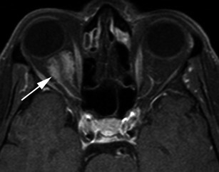
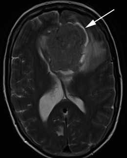
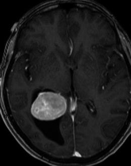
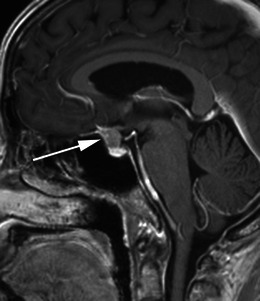
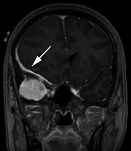
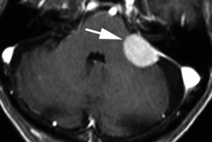
Meningiomas are the most common non-glial tumour of the central nervous system (CNS). There are a number of characteristic imaging features of meningiomas on magnetic resonance imaging (MRI) that allow an accurate diagnosis, however there are a number of atypical features that may be diagnostically challenging. Furthermore, a number of other neoplastic and non-neoplastic conditions may mimic meningiomas. This pictorial review discusses the typical and atypical MRI features of meningiomas and their mimics.
There are several characteristic features of meningiomas on MRI that allow an accurate diagnosis Some meningiomas may display atypical imaging characteristics that may be diagnostically challenging Routine MRI sequences do not reliably distinguish between benign and malignant meningiomas Spectroscopy and diffusion tensor imaging may be useful in the diagnosis of malignant meningiomas A number of conditions may mimic meningiomas; however, they may have additional differentiating features.
脑膜瘤是中枢神经系统(CNS)最常见的非神经胶质肿瘤。脑膜瘤在磁共振成像(MRI)上有许多特征性的影像学表现,可做出准确诊断,但也有一些不典型特征可能具有诊断挑战性。此外,还有一些其他肿瘤性和非肿瘤性疾病可能类似于脑膜瘤。本影像学综述讨论了脑膜瘤及其类似物的典型和非典型 MRI 特征。
MRI 上脑膜瘤有几个特征性表现,有助于准确诊断。一些脑膜瘤可能表现出不典型的影像学特征,可能具有诊断挑战性。常规 MRI 序列不能可靠地区分良性和恶性脑膜瘤。波谱和弥散张量成像可能有助于恶性脑膜瘤的诊断。许多疾病可能类似于脑膜瘤;然而,它们可能有其他有区别的特征。