Department of Physical Therapy and Rehabilitation Science, University of California, San Francisco, CA 94158, USA.
BMC Musculoskelet Disord. 2014 Jan 16;15:19. doi: 10.1186/1471-2474-15-19.
The prevalence of hyperkyphosis is increased in older men; however, risk factors other than age and vertebral fractures are not well established. We previously reported that poor paraspinal muscle composition contributes to more severe kyphosis in a cohort of both older men and women.
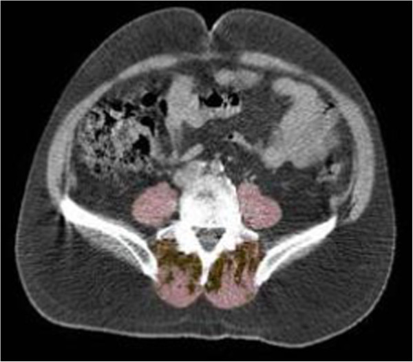
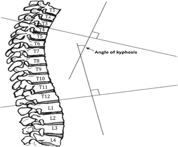
To specifically evaluate this association in older men, we conducted a cross-sectional study to evaluate the association of paraspinal muscle composition and degree of thoracic kyphosis in an analytic cohort of 475 randomly selected participants from the Osteoporotic Fractures in Men (MrOS) study with baseline abdominal quantitative computed tomography (QCT) scans and plain thoracic radiographs. Baseline abdominal QCT scans were used to obtain abdominal body composition measurements of paraspinal muscle and adipose tissue distribution. Supine lateral spine radiographs were used to measure Cobb angle of kyphosis. We examined the linear association of muscle volume, fat volume and kyphosis using loess plots. Multivariate linear models were used to investigate the association between muscle and kyphosis using total muscle volume, as well as individual components of the total muscle volume, including adipose and muscle compartments alone, controlling for age, height, vertebral fractures, and total hip bone mineral density (BMD). We examined these associations among those with no prevalent vertebral fracture and those with BMI < 30 kg/m2.
Among men in the analytic cohort, means (SD) were 74 (SD = 5.9) years for age, and 37.5 (SD = 11.9) degrees for Cobb angle of kyphosis. Men in the lowest tertile of total paraspinal muscle volume had greater mean Cobb angle than men in the highest tertile, although test of linear trend across tertiles did not reach statistical significance. Neither lower paraspinal skeletal muscle volume (p-trend = 0.08), or IMAT (p-trend = 0.96) was associated with greater kyphosis. Results were similar among those with no prevalent vertebral fractures. However, among men with BMI < 30 kg/m2, those in the lowest tertile of paraspinal muscle volume had greater adjusted mean kyphosis (40.0, 95% CI: 37.8 - 42.1) compared to the highest tertile (36.3, 95% CI: 34.2 - 38.4).
These results suggest that differences in body composition may potentially influence kyphosis.
在老年男性中,脊柱后凸的发病率增加;然而,除年龄和椎体骨折外,其他危险因素尚未得到很好的确定。我们之前的研究报告表明,较差的脊柱旁肌肉组成会导致老年男性和女性队列中更严重的后凸。
为了专门评估老年男性的这种相关性,我们进行了一项横断面研究,以评估在来自男性骨质疏松性骨折研究(MrOS)的一个分析队列中,脊柱旁肌肉成分与胸段后凸程度之间的相关性,该队列由 475 名随机选择的参与者组成,基线时有腹部定量计算机断层扫描(QCT)扫描和普通胸部 X 线片。基线腹部 QCT 扫描用于获得脊柱旁肌肉和脂肪组织分布的腹部身体成分测量值。仰卧侧位脊柱 X 射线用于测量后凸的 Cobb 角。我们使用 loess 图检查肌肉体积、脂肪体积和后凸的线性关联。使用总肌肉体积以及总肌肉体积的各个组成部分(包括单独的脂肪和肌肉隔室),控制年龄、身高、椎体骨折和全髋关节骨密度(BMD),使用多元线性模型研究肌肉与后凸之间的关系。我们在没有明显椎体骨折的人群和 BMI<30kg/m2 的人群中检查了这些关联。
在分析队列中的男性中,年龄的平均值(标准差)为 74(标准差=5.9)岁,Cobb 角的平均值(标准差)为 37.5(标准差=11.9)度。总脊柱旁肌肉体积最低三分位的男性平均 Cobb 角大于最高三分位的男性,尽管三分位之间的线性趋势检验未达到统计学意义。较低的脊柱旁骨骼肌体积(p 趋势=0.08)或 IMAT(p 趋势=0.96)与更大的后凸均无关。在没有明显椎体骨折的人群中,结果相似。然而,在 BMI<30kg/m2 的男性中,脊柱旁肌肉体积最低三分位的男性调整后的平均后凸(40.0,95%置信区间:37.8-42.1)大于最高三分位(36.3,95%置信区间:34.2-38.4)。
这些结果表明,身体成分的差异可能会影响后凸。