Mineshige Takayuki, Yasuno Kyohei, Sugahara Go, Tomishita Yoshifumi, Shimokawa Namiko, Kamiie Junichi, Nishifuji Koji, Shirota Kinji
Research Institute of Biosciences, Azabu University, 1-17-71 Fuchinobe, Chuo-ku, Sagamihara, Kanagawa 252-5201, Japan.
J Vet Med Sci. 2014 May;76(5):735-9. doi: 10.1292/jvms.13-0516. Epub 2014 Jan 16.
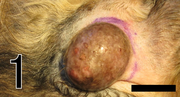
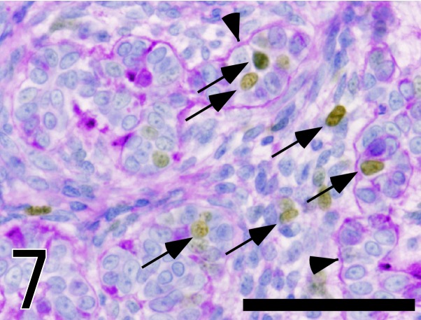
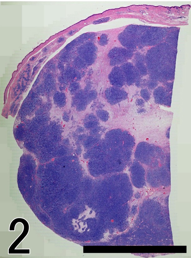
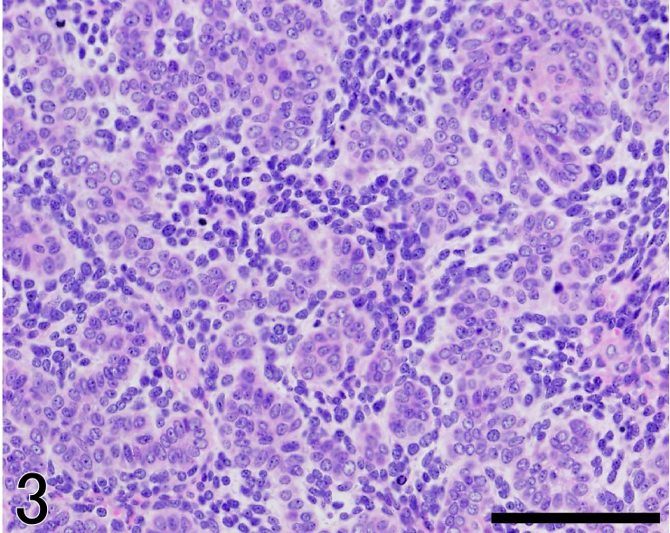
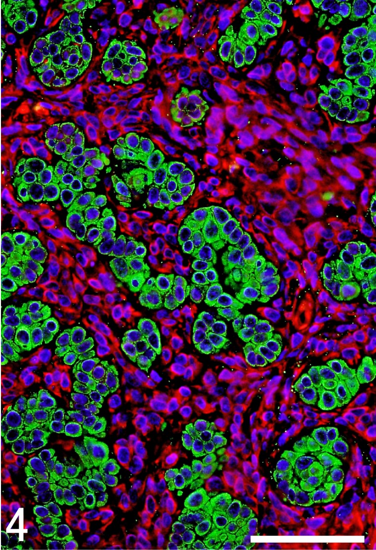
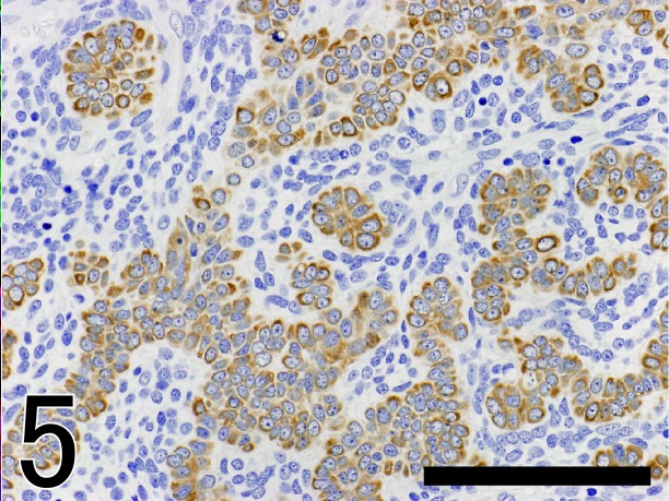
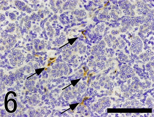
Histopathological and immunohistochemical examinations were made on a cutaneous tumor on the head of an 11-year-old female mixed-breed dog. The tumor was well demarcated and comprised multilobular structures of neoplastic epithelial cells with abundant plump peritumoral stromal cells. The neoplastic cells formed irregular cell cords or trabeculae and were arranged in characteristic palisades at the periphery. Immunohistochemically, neoplastic cells were positive for p63 and the several cytokeratins examined. In contrast, the plump peritumoral stromal cells were positive for vimentin and unevenly for nestin, a neuroepithelial stem cell protein. The stromal cells prominently proliferated in proximity to epithelial neoplastic cells, suggesting a close interaction between these two cell types.
对一只11岁雌性杂种犬头部的皮肤肿瘤进行了组织病理学和免疫组织化学检查。肿瘤边界清晰,由肿瘤上皮细胞的多叶结构组成,周围有丰富的丰满间质细胞。肿瘤细胞形成不规则的细胞索或小梁,并在周边呈特征性栅栏状排列。免疫组织化学显示,肿瘤细胞对p63和所检测的几种细胞角蛋白呈阳性反应。相比之下,丰满的肿瘤周围间质细胞波形蛋白呈阳性,神经上皮干细胞蛋白巢蛋白呈不均匀阳性。间质细胞在肿瘤上皮细胞附近显著增殖,表明这两种细胞类型之间存在密切相互作用。