Palmai Zoltan, Seifert Christian, Gräter Frauke, Balog Erika
Department of Biophysics and Radiation Biology, Semmelweis University, Budapest, Hungary.
Molecular Biomechanics, Heidelberger Institut für Theoretische Studien gGmbH, Heidelberg, Germany.
PLoS Comput Biol. 2014 Jan;10(1):e1003444. doi: 10.1371/journal.pcbi.1003444. Epub 2014 Jan 23.
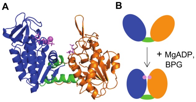
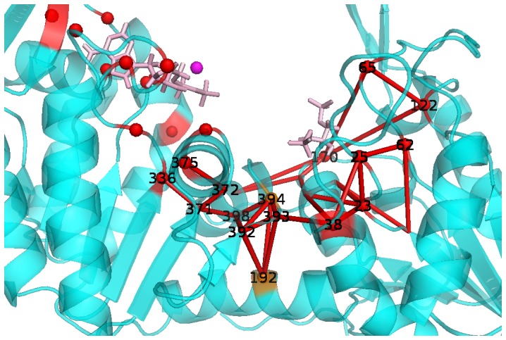
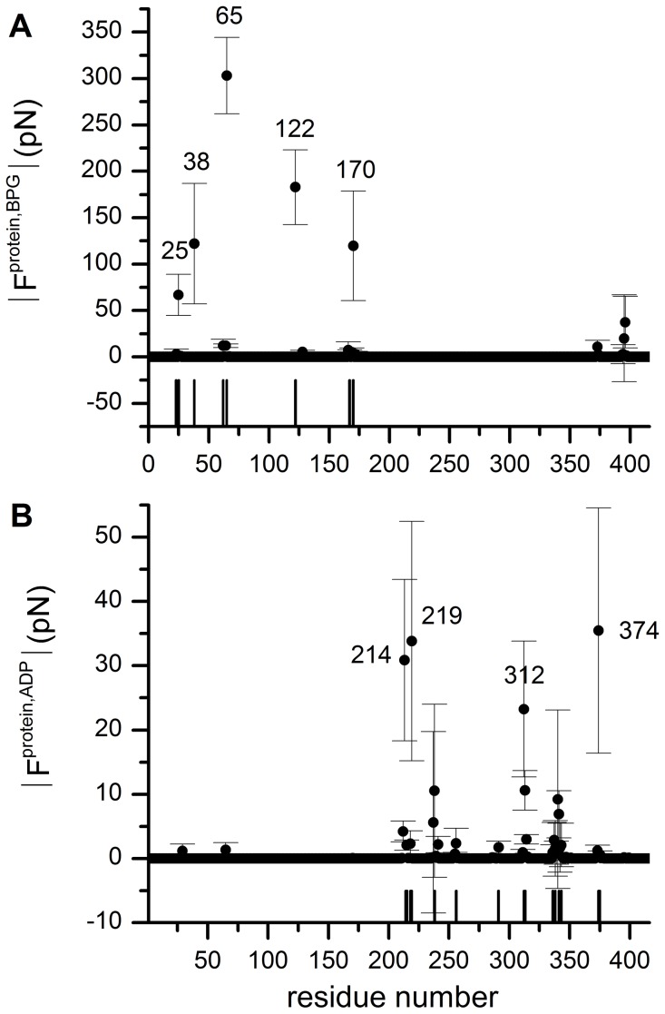
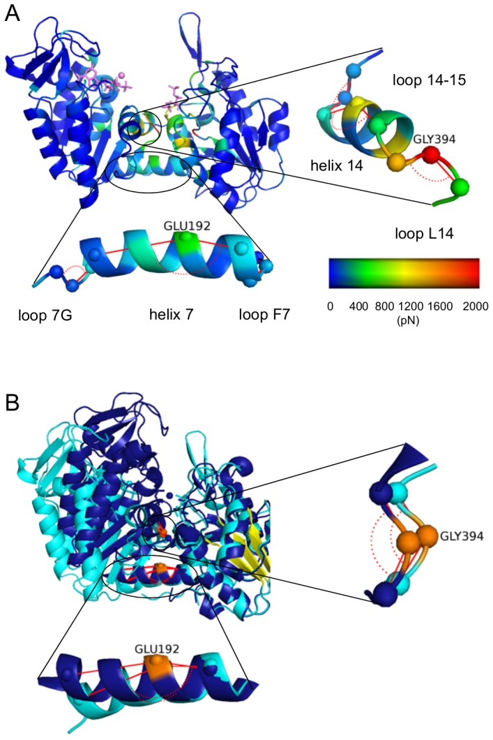
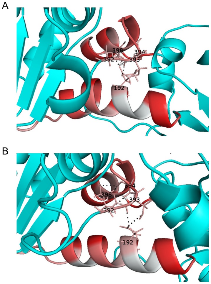
3-Phosphogycerate kinase (PGK) is a two domain enzyme, which transfers a phosphate group between its two substrates, 1,3-bisphosphoglycerate bound to the N-domain and ADP bound to the C-domain. Indispensable for the phosphoryl transfer reaction is a large conformational change from an inactive open to an active closed conformation via a hinge motion that should bring substrates into close proximity. The allosteric pathway resulting in the active closed conformation has only been partially uncovered. Using Molecular Dynamics simulations combined with Force Distribution Analysis (FDA), we describe an allosteric pathway, which connects the substrate binding sites to the interdomain hinge region. Glu192 of alpha-helix 7 and Gly394 of loop L14 act as hinge points, at which these two secondary structure elements straighten, thereby moving the substrate-binding domains towards each other. The long-range allosteric pathway regulating hPGK catalytic activity, which is partially validated and can be further tested by mutagenesis, highlights the virtue of monitoring internal forces to reveal signal propagation, even if only minor conformational distortions, such as helix bending, initiate the large functional rearrangement of the macromolecule.
3-磷酸甘油酸激酶(PGK)是一种双结构域酶,它在其两个底物之间转移磷酸基团,一个底物是结合在N结构域的1,3-二磷酸甘油酸,另一个底物是结合在C结构域的ADP。磷酸转移反应所必需的是通过铰链运动从无活性的开放构象到活性的封闭构象的大的构象变化,这种运动应使底物紧密靠近。导致活性封闭构象的变构途径仅被部分揭示。通过结合分子动力学模拟和力分布分析(FDA),我们描述了一条变构途径,该途径将底物结合位点连接到结构域间的铰链区域。α螺旋7的Glu192和环L14的Gly394作为铰链点,在这些点上这两个二级结构元件伸直,从而使底物结合结构域相互靠近。调节人PGK催化活性的远程变构途径已得到部分验证,可通过诱变进一步测试,这突出了监测内力以揭示信号传播的优点,即使只有微小的构象扭曲,如螺旋弯曲,也能引发大分子的大功能重排。