1. Institute of Functional Nano & Soft Materials (FUNSOM), Soochow University, Suzhou, Jiangsu 215123, China ; 2. Laboratory of Molecular Imaging and Nanomedicine (LOMIN), National Institute of Biomedical Imaging and Bioengineering (NIBIB), National Institutes of Health (NIH), Bethesda, Maryland 20892, United States.
2. Laboratory of Molecular Imaging and Nanomedicine (LOMIN), National Institute of Biomedical Imaging and Bioengineering (NIBIB), National Institutes of Health (NIH), Bethesda, Maryland 20892, United States ; 3. Center for Molecular Imaging and Translational Medicine School of Public Health, Xiamen University, Xiamen 361005, China.
Theranostics. 2014 Jan 2;4(2):134-41. doi: 10.7150/thno.7217. eCollection 2014.
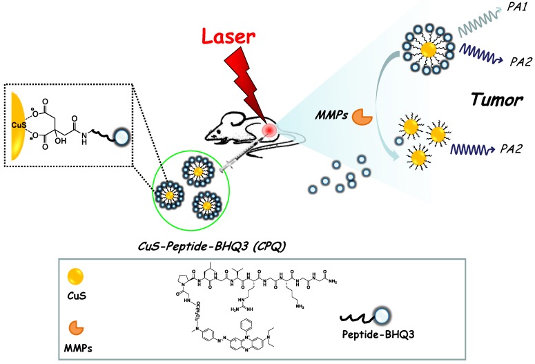
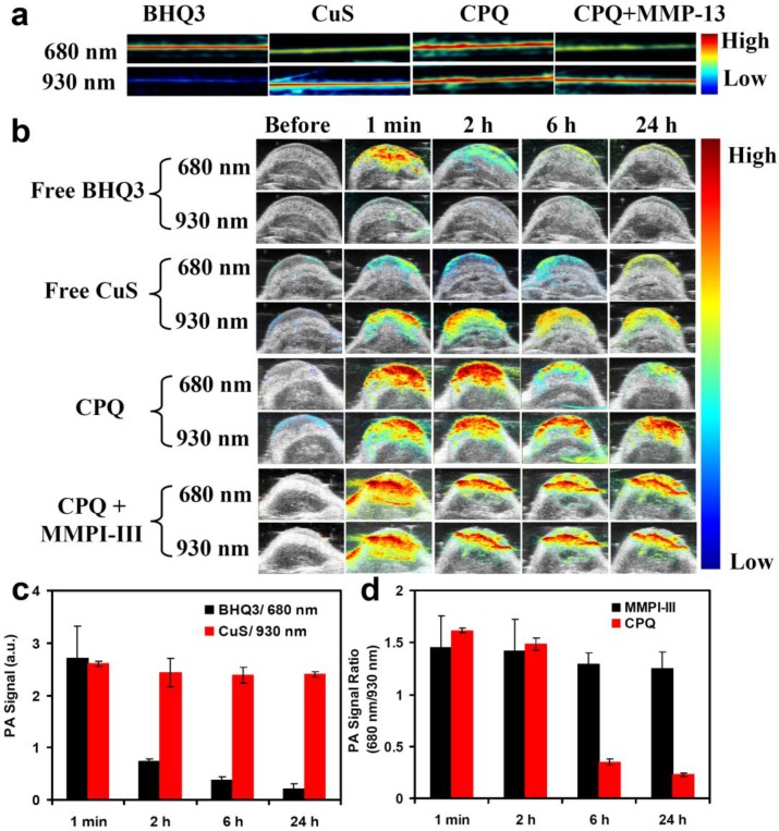
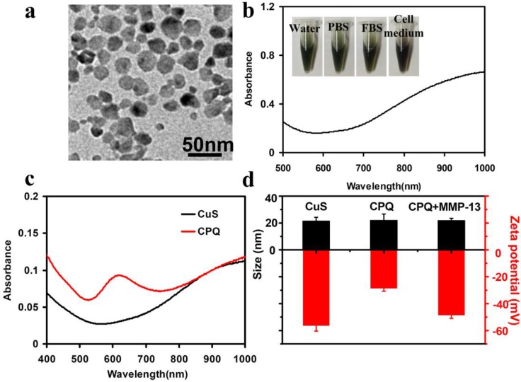
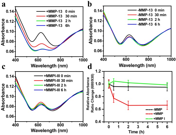
Herein, we for the first time report a novel activatable photoacoustic (PA) imaging nano-probe for in vivo detection of cancer-related matrix metalloproteinases (MMPs). A black hole quencher 3 (BHQ3) which absorbs red light is conjugated to near-infrared (NIR)-absorbing copper sulfide (CuS) nanoparticles via a MMP-cleavable peptide linker. The obtained CuS-peptide-BHQ3 (CPQ) nano-probe exhibits two distinctive absorption peaks at 630 nm and 930 nm. Inside the tumor microenvironment where MMPs present, the MMP-sensitive peptide would be cleaved, releasing BHQ3 from the CuS nanoparticles, the former of which as a small molecule is then rapidly cleared out from the tumor, whereas the latter of which as large nanoparticles would retain inside the tumor for a much longer period of time. As the result, the PA signal at 680 nm which is contributed by BHQ3 would be quickly diminished while that at 930 nm would be largely retained. The PA signal ratio of 680 nm / 930 nm could thus serve as an in vivo indicator of MMPs activity inside the tumor. Our work presents a novel strategy of in vivo sensing of MMPs based on PA imaging, which should offer remarkably improved detection depth compared with traditional optical imaging techniques.
在这里,我们首次报道了一种新型的可激活光声(PA)成像纳米探针,用于体内检测与癌症相关的基质金属蛋白酶(MMPs)。通过 MMP 可切割的肽接头将吸收红光的黑洞猝灭剂 3(BHQ3)与近红外(NIR)吸收的硫化铜(CuS)纳米颗粒连接。所得的 CuS-肽-BHQ3(CPQ)纳米探针在 630nm 和 930nm 处表现出两个独特的吸收峰。在 MMP 存在的肿瘤微环境中,MMP 敏感肽会被切割,从而将 BHQ3 从 CuS 纳米颗粒中释放出来,前者作为小分子会迅速从肿瘤中清除,而后者作为大纳米颗粒会在肿瘤中保留更长的时间。结果,由 BHQ3 贡献的 680nm 的 PA 信号会迅速减弱,而 930nm 的 PA 信号会被大量保留。因此,680nm/930nm 的 PA 信号比可作为肿瘤内 MMPs 活性的体内指示剂。我们的工作提出了一种基于 PA 成像的 MMPs 体内传感的新策略,与传统的光学成像技术相比,它应该提供显著提高的检测深度。