• 文献检索
  • 文档翻译
  • 深度研究
  • 学术资讯
  • Suppr Zotero 插件Zotero 插件
  • 邀请有礼
  • 套餐&价格
  • 历史记录
应用&插件
Suppr Zotero 插件Zotero 插件浏览器插件Mac 客户端Windows 客户端微信小程序
定价
高级版会员购买积分包购买API积分包
服务
文献检索文档翻译深度研究API 文档MCP 服务
关于我们
关于 Suppr公司介绍联系我们用户协议隐私条款
关注我们

Suppr 超能文献

核心技术专利:CN118964589B侵权必究
粤ICP备2023148730 号-1Suppr @ 2026

文献检索

告别复杂PubMed语法,用中文像聊天一样搜索,搜遍4000万医学文献。AI智能推荐,让科研检索更轻松。

立即免费搜索

文件翻译

保留排版,准确专业,支持PDF/Word/PPT等文件格式,支持 12+语言互译。

免费翻译文档

深度研究

AI帮你快速写综述,25分钟生成高质量综述,智能提取关键信息,辅助科研写作。

立即免费体验

使用频谱超声成像对三维工程组织构建体中的体外成骨细胞分化进行无创定量分析。

Noninvasive quantification of in vitro osteoblastic differentiation in 3D engineered tissue constructs using spectral ultrasound imaging.

作者信息

Gudur Madhu Sudhan Reddy, Rao Rameshwar R, Peterson Alexis W, Caldwell David J, Stegemann Jan P, Deng Cheri X

机构信息

Department of Biomedical Engineering, University of Michigan, Ann Arbor, Michigan, United States of America.

出版信息

PLoS One. 2014 Jan 22;9(1):e85749. doi: 10.1371/journal.pone.0085749. eCollection 2014.

DOI:10.1371/journal.pone.0085749
PMID:24465680
原文链接:https://pmc.ncbi.nlm.nih.gov/articles/PMC3899074/
Abstract

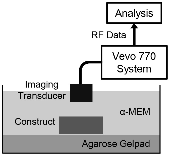
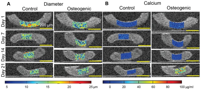
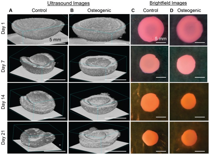
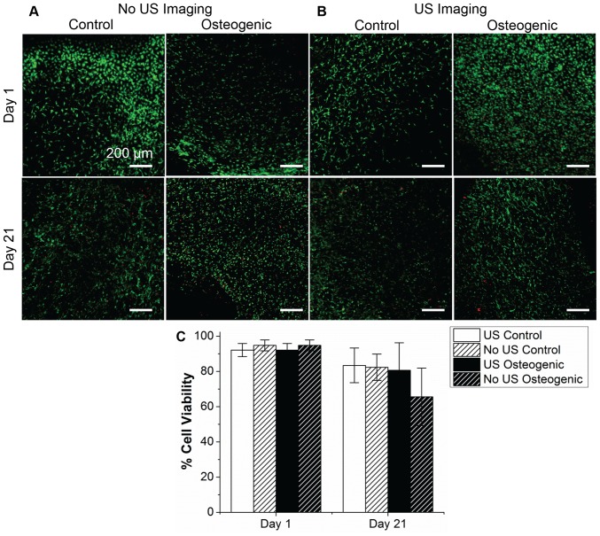
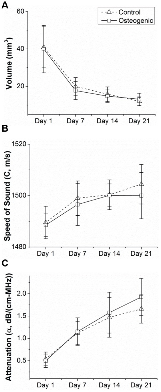
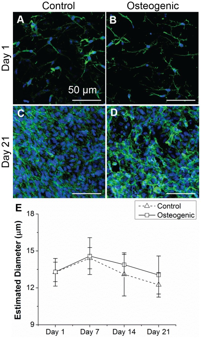
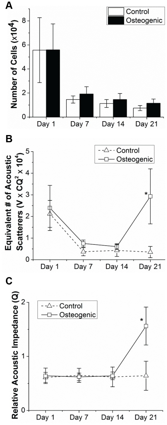
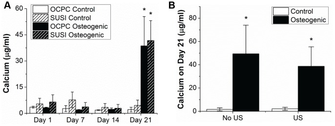
Non-destructive monitoring of engineered tissues is needed for translation of these products from the lab to the clinic. In this study, non-invasive, high resolution spectral ultrasound imaging (SUSI) was used to monitor the differentiation of MC3T3 pre-osteoblasts seeded within collagen hydrogels. SUSI was used to measure the diameter, concentration and acoustic attenuation of scatterers within such constructs cultured in either control or osteogenic medium over 21 days. Conventional biochemical assays were used on parallel samples to determine DNA content and calcium deposition. Construct volume and morphology were accurately imaged using ultrasound. Cell diameter was estimated to be approximately 12.5-15.5 µm using SUSI, which corresponded well to measurements of fluorescently stained cells. The total number of cells per construct assessed by quantitation of DNA content decreased from 5.6±2.4×10(4) at day 1 to 0.9±0.2×10(4) at day 21. SUSI estimation of the equivalent number of acoustic scatters showed a similar decreasing trend, except at day 21 in the osteogenic samples, which showed a marked increase in both scatterer number and acoustic impedance, suggestive of mineral deposition by the differentiating MC3T3 cells. Estimation of calcium content by SUSI was 41.7±11.4 µg/ml, which agreed well with the biochemical measurement of 38.7±16.7 µg/ml. Color coded maps of parameter values were overlaid on B-mode images to show spatiotemporal changes in cell diameter and calcium deposition. This study demonstrates the use of non-destructive ultrasound imaging to provide quantitative information on the number and differentiated state of cells embedded within 3D engineered constructs, and therefore presents a valuable tool for longitudinal monitoring of engineered tissue development.

摘要

为了将这些产品从实验室转化到临床,需要对工程组织进行无损监测。在本研究中,使用非侵入性高分辨率光谱超声成像(SUSI)监测接种在胶原水凝胶中的MC3T3前成骨细胞的分化。SUSI用于测量在对照或成骨培养基中培养21天的此类构建体中散射体的直径、浓度和声衰减。对平行样本进行传统生化分析以确定DNA含量和钙沉积。使用超声准确成像构建体的体积和形态。使用SUSI估计细胞直径约为12.5 - 15.5 µm,这与荧光染色细胞的测量结果非常吻合。通过DNA含量定量评估的每个构建体的细胞总数从第1天的5.6±2.4×10⁴减少到第21天的0.9±0.2×10⁴。SUSI对等效声散射体数量的估计显示出类似的下降趋势,但在成骨样本的第21天除外,此时散射体数量和声阻抗均显著增加,提示分化的MC3T3细胞发生了矿物质沉积。SUSI估计的钙含量为41.7±11.4 µg/ml,与生化测量值38.7±16.7 µg/ml非常吻合。参数值的彩色编码图叠加在B模式图像上,以显示细胞直径和钙沉积的时空变化。本研究证明了使用无损超声成像来提供关于嵌入三维工程构建体中细胞数量和分化状态的定量信息,因此为工程组织发育的纵向监测提供了一个有价值的工具。

https://cdn.ncbi.nlm.nih.gov/pmc/blobs/9929/3899074/5edcc5c8e328/pone.0085749.g008.jpg
https://cdn.ncbi.nlm.nih.gov/pmc/blobs/9929/3899074/e4098885ed5d/pone.0085749.g001.jpg
https://cdn.ncbi.nlm.nih.gov/pmc/blobs/9929/3899074/3a71b46c7b2a/pone.0085749.g002.jpg
https://cdn.ncbi.nlm.nih.gov/pmc/blobs/9929/3899074/af6a6ed73357/pone.0085749.g003.jpg
https://cdn.ncbi.nlm.nih.gov/pmc/blobs/9929/3899074/aec4cc8f13f9/pone.0085749.g004.jpg
https://cdn.ncbi.nlm.nih.gov/pmc/blobs/9929/3899074/94ed192bcdd6/pone.0085749.g005.jpg
https://cdn.ncbi.nlm.nih.gov/pmc/blobs/9929/3899074/9696fa721cf8/pone.0085749.g006.jpg
https://cdn.ncbi.nlm.nih.gov/pmc/blobs/9929/3899074/6bbe3fced8cb/pone.0085749.g007.jpg
https://cdn.ncbi.nlm.nih.gov/pmc/blobs/9929/3899074/5edcc5c8e328/pone.0085749.g008.jpg

相似文献

1
Noninvasive quantification of in vitro osteoblastic differentiation in 3D engineered tissue constructs using spectral ultrasound imaging.使用频谱超声成像对三维工程组织构建体中的体外成骨细胞分化进行无创定量分析。
PLoS One. 2014 Jan 22;9(1):e85749. doi: 10.1371/journal.pone.0085749. eCollection 2014.
2
Noninvasive, quantitative, spatiotemporal characterization of mineralization in three-dimensional collagen hydrogels using high-resolution spectral ultrasound imaging.使用高分辨率光谱超声成像技术无创、定量、时空地描绘三维胶原水凝胶中的矿化。
Tissue Eng Part C Methods. 2012 Dec;18(12):935-46. doi: 10.1089/ten.TEC.2012.0180. Epub 2012 Jul 16.
3
Non-invasive time-lapsed monitoring and quantification of engineered bone-like tissue.工程化骨样组织的非侵入性实时监测与定量分析
Ann Biomed Eng. 2007 Oct;35(10):1657-67. doi: 10.1007/s10439-007-9338-2. Epub 2007 Jun 2.
4
Complex permittivity measurement as a new noninvasive tool for monitoring in vitro tissue engineering and cell signature through the detection of cell proliferation, differentiation, and pretissue formation.复介电常数测量作为一种新型非侵入性工具,通过检测细胞增殖、分化和组织前体形成来监测体外组织工程和细胞特征。
IEEE Trans Nanobioscience. 2004 Dec;3(4):243-50. doi: 10.1109/tnb.2004.837901.
5
Polymeric Microspheres/Cells/Extracellular Matrix Constructs Produced by Auto-Assembly for Bone Modular Tissue Engineering.自组装制备用于骨组织工程模块化的聚合微球/细胞/细胞外基质构建体。
Int J Mol Sci. 2021 Jul 23;22(15):7897. doi: 10.3390/ijms22157897.
6
[Rotating three-dimensional dynamic culture of osteoblasts seeded on segmental scaffolds with controlled internal channel architectures for construction of segmental tissue engineered bone in vitro].[基于具有可控内部通道结构的节段性支架上接种成骨细胞的旋转三维动态培养用于体外构建节段性组织工程骨]
Zhonghua Yi Xue Za Zhi. 2007 Jan 16;87(3):200-3.
7
Osteogenic differentiation of human periosteal-derived cells in a three-dimensional collagen scaffold.人骨膜来源细胞在三维胶原支架中的成骨分化。
Mol Biol Rep. 2011 Jun;38(5):2887-94. doi: 10.1007/s11033-010-9950-3. Epub 2010 Jan 28.
8
Study of osteoblastic cells in a microfluidic environment.微流控环境中骨细胞的研究。
Biomaterials. 2006 Feb;27(4):586-95. doi: 10.1016/j.biomaterials.2005.06.002. Epub 2005 Jul 18.
9
Runx2/Cbfa1-genetically engineered skeletal myoblasts mineralize collagen scaffolds in vitro.经基因工程改造的Runx2/Cbfa1骨骼肌成肌细胞在体外可使胶原支架矿化。
Biotechnol Bioeng. 2004 Nov 5;88(3):369-78. doi: 10.1002/bit.20251.
10
Effect of bone extracellular matrix synthesized in vitro on the osteoblastic differentiation of marrow stromal cells.体外合成的骨细胞外基质对骨髓基质细胞成骨分化的影响
Biomaterials. 2005 Mar;26(9):971-7. doi: 10.1016/j.biomaterials.2004.04.001.

引用本文的文献

1
New Nanobioceramics Based on Hydroxyapatite for Biomedical Applications: Stability and Properties.用于生物医学应用的基于羟基磷灰石的新型纳米生物陶瓷:稳定性与性能
Nanomaterials (Basel). 2025 Jan 30;15(3):224. doi: 10.3390/nano15030224.
2
Digital Light Processing of F MRI-Traceable Gelatin-Based Biomaterial Inks towards Bone Tissue Regeneration.用于骨组织再生的功能磁共振成像可追踪的明胶基生物材料墨水的数字光处理
Materials (Basel). 2024 Jun 19;17(12):2996. doi: 10.3390/ma17122996.
3
Echo intensity and gray-level co-occurrence matrix analysis of soft tissue grafting biomaterials and dental implants: an ultrasonographic pilot study.

本文引用的文献

1
Winner for outstanding research in the Ph.D. category for the 2013 Society for Biomaterials meeting and exposition, April 10-13, 2013, Boston, Massachusetts: Osteogenic differentiation of adipose-derived and marrow-derived mesenchymal stem cells in modular protein/ceramic microbeads.2013 年生物材料学会会议和博览会博士组杰出研究奖获得者,2013 年 4 月 10 日-13 日,马萨诸塞州波士顿:模块化蛋白/陶瓷微珠中脂肪来源和骨髓来源间充质干细胞的成骨分化。
J Biomed Mater Res A. 2013 Jun;101(6):1531-8. doi: 10.1002/jbm.a.34611. Epub 2013 Mar 30.
2
High-frequency rapid B-mode ultrasound imaging for real-time monitoring of lesion formation and gas body activity during high-intensity focused ultrasound ablation.高频快速 B 模式超声成像用于实时监测高强度聚焦超声消融过程中病变形成和气体的活性。
IEEE Trans Ultrason Ferroelectr Freq Control. 2012 Aug;59(8):1687-99. doi: 10.1109/TUFFC.2012.2374.
3
软组织移植生物材料和牙种植体的超声回声强度和灰度共生矩阵分析:一项超声初步研究。
Dentomaxillofac Radiol. 2023 Oct;52(7):20230033. doi: 10.1259/dmfr.20230033. Epub 2023 Jul 4.
4
Biological and Physico-Chemical Properties of Composite Layers Based on Magnesium-Doped Hydroxyapatite in Chitosan Matrix.基于壳聚糖基质中镁掺杂羟基磷灰石的复合层的生物学和物理化学性质
Micromachines (Basel). 2022 Sep 22;13(10):1574. doi: 10.3390/mi13101574.
5
Therapeutic Ultrasound Effects on Human Induced Pluripotent Stem Cell Cardiomyocytes Measured Optically and with Spectral Ultrasound.光学和超声光谱法测量人诱导多能干细胞心肌细胞的治疗超声效应。
Ultrasound Med Biol. 2022 Jun;48(6):1078-1094. doi: 10.1016/j.ultrasmedbio.2022.02.006. Epub 2022 Mar 15.
6
A novel quantitative and reference-free ultrasound analysis to discriminate different concentrations of bone mineral content.一种新型的定量且无需参考标准的超声分析方法,用于区分不同浓度的骨矿物质含量。
Sci Rep. 2021 Jan 11;11(1):301. doi: 10.1038/s41598-020-79365-0.
7
Multimodal Label-Free Imaging for Detecting Maturation of Engineered Osteogenic Grafts.用于检测工程化骨生成移植物成熟度的多模态无标记成像
ACS Biomater Sci Eng. 2019 Apr 8;5(4):1956-1966. doi: 10.1021/acsbiomaterials.9b00007. Epub 2019 Mar 27.
8
Injectable pre-cultured tissue modules catalyze the formation of extensive functional microvasculature in vivo.可注射预培养组织模块在体内催化广泛功能性微血管的形成。
Sci Rep. 2020 Sep 23;10(1):15562. doi: 10.1038/s41598-020-72576-5.
9
Photoacoustic Imaging in Tissue Engineering and Regenerative Medicine.组织工程与再生医学中的光声成像。
Tissue Eng Part B Rev. 2020 Feb;26(1):79-102. doi: 10.1089/ten.TEB.2019.0296. Epub 2020 Jan 14.
10
High Frequency Spectral Ultrasound Imaging to Detect Metastasis in Implanted Biomaterial Scaffolds.高频超声光谱成像检测植入生物材料支架中的转移。
Ann Biomed Eng. 2020 Jan;48(1):477-489. doi: 10.1007/s10439-019-02366-2. Epub 2019 Sep 23.
Measuring bone mineral density with fat-water MRI: comparison with computed tomography.利用脂肪水 MRI 测量骨密度:与计算机断层扫描的比较。
J Magn Reson Imaging. 2013 Jan;37(1):237-42. doi: 10.1002/jmri.23749. Epub 2012 Jul 10.
4
In search of an osteoblast cell model for in vitro research.寻找用于体外研究的成骨细胞细胞模型。
Eur Cell Mater. 2012 Jul 9;24:1-17. doi: 10.22203/ecm.v024a01.
5
Noninvasive, quantitative, spatiotemporal characterization of mineralization in three-dimensional collagen hydrogels using high-resolution spectral ultrasound imaging.使用高分辨率光谱超声成像技术无创、定量、时空地描绘三维胶原水凝胶中的矿化。
Tissue Eng Part C Methods. 2012 Dec;18(12):935-46. doi: 10.1089/ten.TEC.2012.0180. Epub 2012 Jul 16.
6
Characterization of the pancreas in vivo using EUS spectrum analysis with electronic array echoendoscopes.使用电子相控阵超声内镜进行超声谱分析对活体胰腺进行特征描述。
Gastrointest Endosc. 2012 Jun;75(6):1175-83. doi: 10.1016/j.gie.2012.01.039. Epub 2012 Apr 11.
7
Matrix composition regulates three-dimensional network formation by endothelial cells and mesenchymal stem cells in collagen/fibrin materials.基质组成调控内皮细胞和间充质干细胞在胶原/纤维蛋白材料中的三维网络形成。
Angiogenesis. 2012 Jun;15(2):253-64. doi: 10.1007/s10456-012-9257-1. Epub 2012 Mar 2.
8
High-frequency ultrasound m-mode imaging for identifying lesion and bubble activity during high-intensity focused ultrasound ablation.高频超声 M 模式成像在高强度聚焦超声消融中识别病变和气泡活动。
Ultrasound Med Biol. 2012 Apr;38(4):626-41. doi: 10.1016/j.ultrasmedbio.2012.01.004. Epub 2012 Feb 15.
9
Nondestructive evaluation of hydrogel mechanical properties using ultrasound.利用超声波对水凝胶力学性能进行无损评估。
Ann Biomed Eng. 2011 Oct;39(10):2521-30. doi: 10.1007/s10439-011-0351-0. Epub 2011 Jul 20.
10
Nondestructive method to evaluate the collagen content of fibrin-based tissue engineered structures via ultrasound.通过超声评估基于纤维蛋白的组织工程结构中胶原含量的非破坏性方法。
Tissue Eng Part C Methods. 2011 Oct;17(10):1021-6. doi: 10.1089/ten.TEC.2010.0669. Epub 2011 Jul 26.