Shigematsu Hiroaki, Kumagai Kenichi, Kobayashi Hiroshi, Eguchi Takanori, Kitaura Kazutaka, Suzuki Satsuki, Horikawa Tatsuya, Matsutani Takaji, Ogasawara Kouetsu, Hamada Yoshiki, Suzuki Ryuji
Department of Rheumatology and Clinical Immunology, Clinical Research Center for Rheumatology and Allergy, Sagamihara National Hospital, National Hospital Organization, Sagamihara, Japan ; Department of Oral and Maxillofacial Surgery, School of Dental Medicine, Tsurumi University, Yokohama, Japan.
Department of Rheumatology and Clinical Immunology, Clinical Research Center for Rheumatology and Allergy, Sagamihara National Hospital, National Hospital Organization, Sagamihara, Japan ; Department of Oral and Maxillofacial Surgery, School of Dental Medicine, Tsurumi University, Yokohama, Japan ; Department of Oral and Maxillofacial Surgery, Nagano Matsushiro General Hospital, Nagano, Japan.
PLoS One. 2014 Jan 20;9(1):e85983. doi: 10.1371/journal.pone.0085983. eCollection 2014.
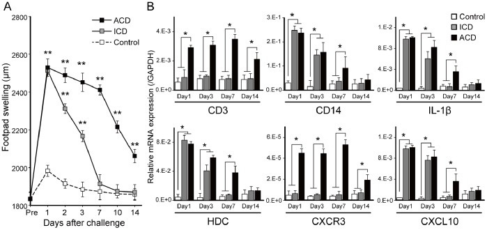
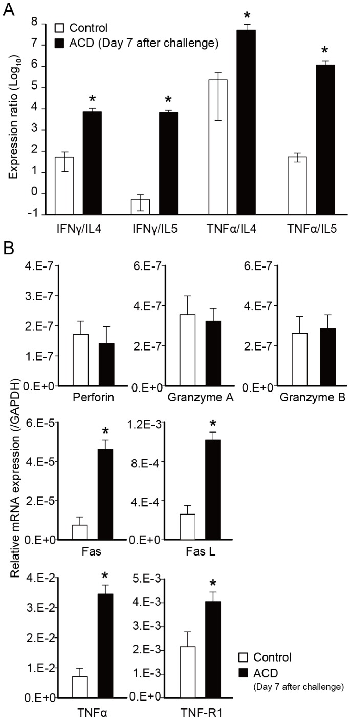
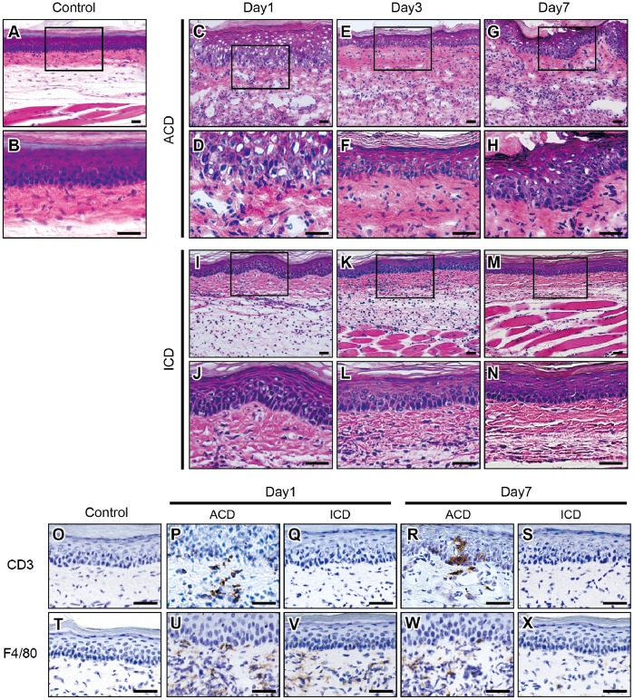
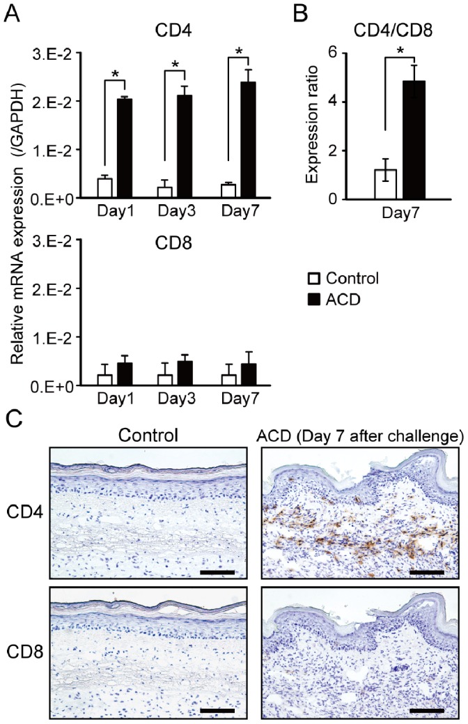
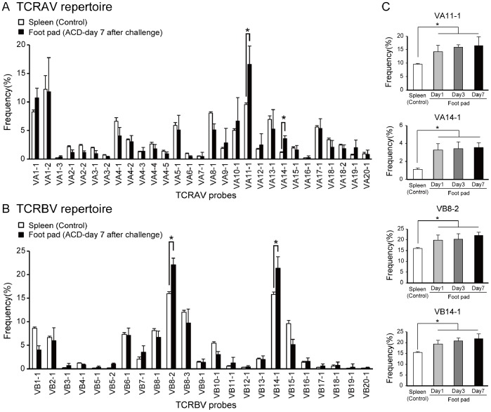
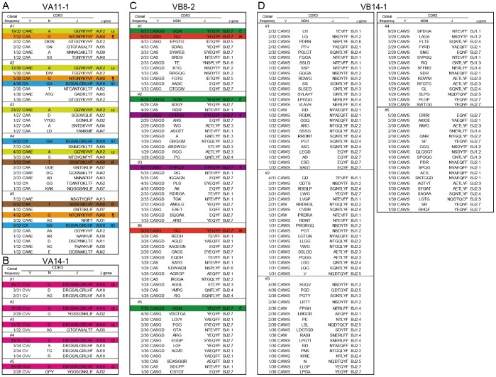
Chromium (Cr) causes delayed-type hypersensitivity reactions possibly mediated by accumulating T cells into allergic inflamed skin, which are called irritants or allergic contact dermatitis. However, accumulating T cells during development of metal allergy are poorly characterized because a suitable animal model is not available. This study aimed to elucidate the skewing of T-cell receptor (TCR) repertoire and cytokine profiles in accumulated T cells in inflamed skin during elucidation of Cr allergy. A novel model of Cr allergy was induced by two sensitizations of Cr plus lipopolysaccharide solution into mouse groin followed by single Cr challenge into the footpad. TCR repertoires and nucleotide sequences of complementary determining region 3 were assessed in accumulated T cells from inflamed skin. Cytokine expression profiles and T-cell phenotypes were determined by qPCR. CD3+CD4+ T cells accumulated in allergic footpads and produced increased T helper 1 (Th1) type cytokines, Fas, and Fas ligand in the footpads after challenge, suggesting CD4+ Th1 cells locally expanded in response to Cr. Accumulated T cells included natural killer (NK) T cells and Cr-specific T cells with VA11-1/VB14-1 usage, suggesting metal-specific T cells driven by invariant NKT cells might contribute to the pathogenesis of Cr allergy.
铬(Cr)可引发迟发型超敏反应,可能是通过T细胞在过敏性炎症皮肤中积聚介导的,这类反应被称为刺激性或过敏性接触性皮炎。然而,由于缺乏合适的动物模型,金属过敏发生过程中T细胞的积聚情况尚未得到充分表征。本研究旨在阐明铬过敏过程中炎症皮肤中积聚的T细胞的T细胞受体(TCR)库和细胞因子谱的偏移情况。通过将铬加脂多糖溶液对小鼠腹股沟进行两次致敏,随后对足垫进行单次铬激发,诱导出一种新型的铬过敏模型。对炎症皮肤中积聚的T细胞的TCR库和互补决定区3的核苷酸序列进行评估。通过qPCR确定细胞因子表达谱和T细胞表型。在激发后,CD3 + CD4 + T细胞在过敏性足垫中积聚,并在足垫中产生增加的辅助性T细胞1(Th1)型细胞因子、Fas和Fas配体,表明CD4 + Th1细胞因铬而在局部扩增。积聚的T细胞包括自然杀伤(NK)T细胞和使用VA11 - 1/VB14 - 1的铬特异性T细胞,这表明由不变自然杀伤T细胞驱动的金属特异性T细胞可能参与铬过敏的发病机制。