Department of Oral and Maxillofacial Surgery, School of Dental Medicine, Tsurumi University, Yokohama, Japan ; Department of Rheumatology and Clinical Immunology, Clinical Research Center for Rheumatology and Allergy, Sagamihara National Hospital, National Hospital Organization, Sagamihara, Japan ; Department of Oral and Maxillofacial Surgery, Nagano Matsushiro General Hospital, Nagano, Japan.
PLoS One. 2013 Oct 3;8(10):e76385. doi: 10.1371/journal.pone.0076385. eCollection 2013.
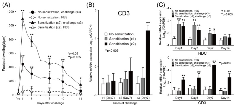
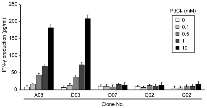
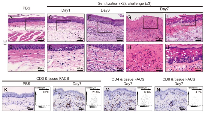
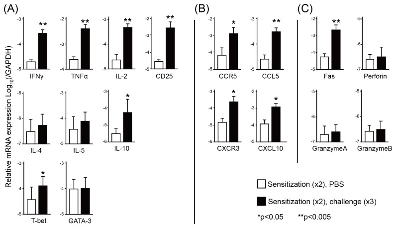
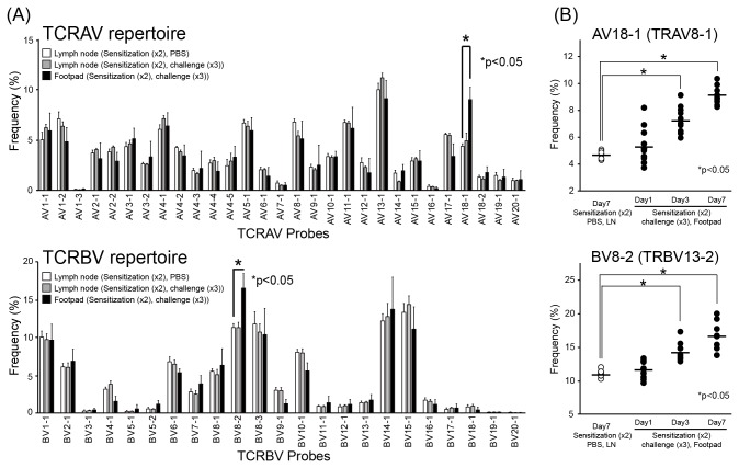
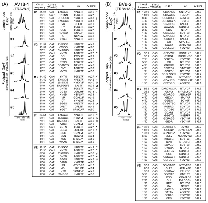
Metal allergy is categorized as a delayed-type hypersensitivity reaction, and is characterized by the recruitment of lymphocytes into sites of allergic inflammation. Because of the unavailability of suitable animal models for metal allergy, the role of T cells in the pathogenesis of metal allergy has not been explored. Thus, we developed a novel mouse model for metal allergy associated with infiltration of T cells by multiple injections of palladium (Pd) plus lipopolysaccharide into the footpad. Using this model, we characterized footpad-infiltrating T cells in terms of phenotypic markers, T cell receptor (TCR) repertoires and cytokine expression. CD3+ CD4+ T cells accumulated in the allergic footpads 7 days after Pd challenge. The expression levels of CD25, interleukin-2, interferon-γ and tumor necrosis factor, but not interleukin-4 and interleukin-5, increased in the footpads after challenge, suggesting CD4+ T helper 1 (Th1) cells locally expanded in response to Pd. Infiltrated T cells in the footpads frequently expressed AV18-1 and BV8-2 T cell receptor (TCR) chains compared with T cells in the lymph nodes and exhibited oligoclonality. T-cell clones identified from Pd-allergic mouse footpads shared identical CDR3 sequences containing AV18-1 and BV8-2. These results suggest that TCR AV18-1 and BV8-2 play dominant and critical parts in the antigen specificity of Pd-specific Th1 cells.
金属过敏被归类为迟发型超敏反应,其特征是淋巴细胞募集到过敏炎症部位。由于缺乏用于金属过敏的合适动物模型,因此尚未探讨 T 细胞在金属过敏发病机制中的作用。因此,我们通过将钯(Pd)和脂多糖多次注射到脚掌中来开发一种与 T 细胞浸润相关的新型金属过敏小鼠模型。使用该模型,我们根据表型标志物、T 细胞受体(TCR)谱和细胞因子表达来表征脚掌浸润的 T 细胞。在 Pd 挑战后 7 天,CD3+CD4+T 细胞在过敏的脚掌中积累。挑战后,脚掌中的 CD25、白细胞介素-2、干扰素-γ和肿瘤坏死因子的表达水平增加,但白细胞介素-4 和白细胞介素-5 的表达水平没有增加,这表明 CD4+T 辅助 1(Th1)细胞对 Pd 产生局部扩增。与淋巴结中的 T 细胞相比,脚掌中浸润的 T 细胞经常表达 AV18-1 和 BV8-2 TCR 链,并表现出寡克隆性。从 Pd 过敏小鼠脚掌中鉴定出的 T 细胞克隆共享包含 AV18-1 和 BV8-2 的相同 CDR3 序列。这些结果表明,TCR AV18-1 和 BV8-2 在 Pd 特异性 Th1 细胞的抗原特异性中起主要和关键作用。