Chollet Madeleine B, Aldridge Kristina, Pangborn Nicole, Weinberg Seth M, Deleon Valerie B
Center for Functional Anatomy and Evolution, Johns Hopkins University School of Medicine, Baltimore, Maryland, United States of America.
Department of Pathology and Anatomical Sciences, University of Missouri School of Medicine, Columbia, Missouri, United States of America.
PLoS One. 2014 Jan 28;9(1):e86005. doi: 10.1371/journal.pone.0086005. eCollection 2014.
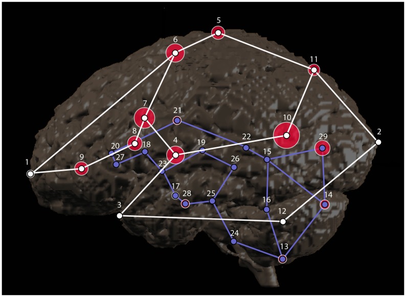
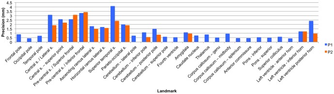
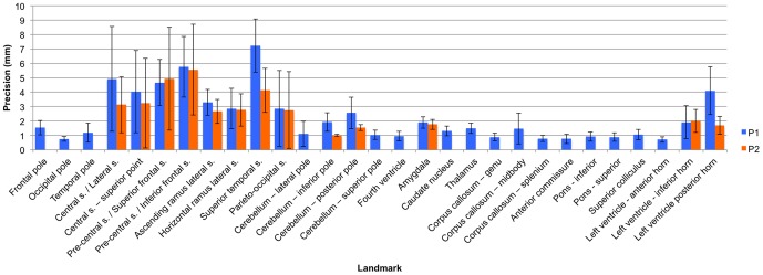
Neuroanatomic phenotypes are often assessed using volumetric analysis. Although powerful and versatile, this approach is limited in that it is unable to quantify changes in shape, to describe how regions are interrelated, or to determine whether changes in size are global or local. Statistical shape analysis using coordinate data from biologically relevant landmarks is the preferred method for testing these aspects of phenotype. To date, approximately fifty landmarks have been used to study brain shape. Of the studies that have used landmark-based statistical shape analysis of the brain, most have not published protocols for landmark identification or the results of reliability studies on these landmarks. The primary aims of this study were two-fold: (1) to collaboratively develop detailed data collection protocols for a set of brain landmarks, and (2) to complete an intra- and inter-observer validation study of the set of landmarks. Detailed protocols were developed for 29 cortical and subcortical landmarks using a sample of 10 boys aged 12 years old. Average intra-observer error for the final set of landmarks was 1.9 mm with a range of 0.72 mm-5.6 mm. Average inter-observer error was 1.1 mm with a range of 0.40 mm-3.4 mm. This study successfully establishes landmark protocols with a minimal level of error that can be used by other researchers in the assessment of neuroanatomic phenotypes.
神经解剖学表型通常采用体积分析进行评估。尽管这种方法功能强大且用途广泛,但它也有局限性,即无法量化形状变化、描述区域之间的相互关系,或者确定大小变化是全局性的还是局部性的。使用来自生物学相关标志点的坐标数据进行统计形状分析是测试表型这些方面的首选方法。迄今为止,大约五十个标志点已被用于研究脑形状。在那些对大脑进行基于标志点的统计形状分析的研究中,大多数都没有公布标志点识别的方案或这些标志点可靠性研究的结果。本研究的主要目的有两个:(1)合作制定一套脑标志点的详细数据收集方案,(2)完成该套标志点的观察者内和观察者间验证研究。使用10名12岁男孩的样本为29个皮质和皮质下标志点制定了详细方案。最终标志点集的观察者内平均误差为1.9毫米,范围为0.72毫米至5.6毫米。观察者间平均误差为1.1毫米,范围为0.40毫米至3.4毫米。本研究成功建立了误差极小的标志点方案,可供其他研究人员用于评估神经解剖学表型。