Paquette N, Shi J, Wang Y, Lao Y, Ceschin R, Nelson M D, Panigrahy A, Lepore N
Department of Radiology, University of Southern California and Children's Hospital of Los Angeles, CA, USA.
School of Computing, Informatics, and Decision Systems Engineering, Arizona State University, Tempe, AZ, USA.
Neuroimage Clin. 2017 May 28;15:483-493. doi: 10.1016/j.nicl.2017.05.025. eCollection 2017.
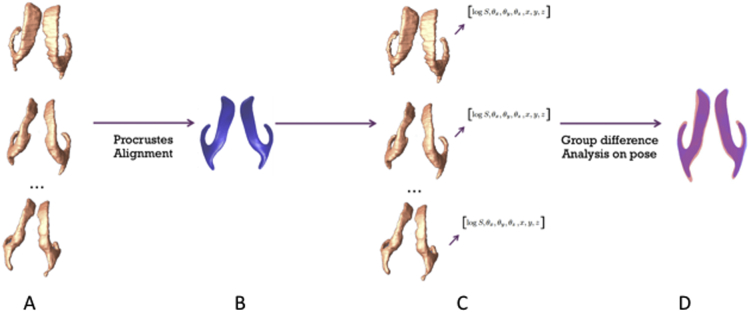
Recent neuroimaging findings have highlighted the impact of premature birth on subcortical development and morphological changes in the deep grey nuclei and ventricular system. To help characterize subcortical microstructural changes in preterm neonates, we recently implemented a multivariate tensor-based method (mTBM). This method allows to precisely measure local surface deformation of brain structures in infants. Here, we investigated ventricular abnormalities and their spatial relationships with surrounding subcortical structures in preterm neonates. We performed regional group comparisons on the surface morphometry and relative position of the lateral ventricles between 19 full-term and 17 preterm born neonates at term-equivalent age. Furthermore, a relative pose analysis was used to detect individual differences in translation, rotation, and scale of a given brain structure with respect to an average. Our mTBM results revealed broad areas of alterations on the frontal horn and body of the left ventricle, and narrower areas of differences on the temporal horn of the right ventricle. A significant shift in the rotation of the left ventricle was also found in preterm neonates. Furthermore, we located significant correlations between morphology and pose parameters of the lateral ventricles and that of the putamen and thalamus. These results show that regional abnormalities on the surface and pose of the ventricles are also associated with alterations on the putamen and thalamus. The complementarity of the information provided by the surface and pose analysis may help to identify abnormal white and grey matter growth, hinting toward a pattern of neural and cellular dysmaturation.
近期的神经影像学研究结果凸显了早产对皮层下发育以及深部灰质核团和脑室系统形态变化的影响。为了帮助刻画早产儿皮层下微观结构的变化,我们最近采用了一种基于张量的多变量方法(mTBM)。这种方法能够精确测量婴儿脑结构的局部表面变形。在此,我们研究了早产儿的脑室异常及其与周围皮层下结构的空间关系。我们对19名足月新生儿和17名足月等效年龄的早产儿的侧脑室表面形态测量和相对位置进行了区域组间比较。此外,我们使用相对位姿分析来检测给定脑结构相对于平均值在平移、旋转和缩放方面的个体差异。我们的mTBM结果显示,左脑室额角和体部存在广泛的改变区域,右脑室颞角的差异区域较窄。在早产儿中还发现左脑室旋转有显著变化。此外,我们发现侧脑室与壳核和丘脑的形态及位姿参数之间存在显著相关性。这些结果表明,脑室表面和位姿的区域异常也与壳核和丘脑的改变有关。表面和位姿分析所提供信息的互补性可能有助于识别白质和灰质的异常生长,提示神经和细胞发育异常的模式。