Queensland Brain Institute, Clem Jones Centre for Ageing Dementia Research, The University of Queensland, Brisbane, Queensland, Australia ; School of Mathematics and Physics, The University of Queensland, Brisbane, Queensland, Australia.
Queensland Brain Institute, Clem Jones Centre for Ageing Dementia Research, The University of Queensland, Brisbane, Queensland, Australia.
PLoS One. 2014 Jan 29;9(1):e87242. doi: 10.1371/journal.pone.0087242. eCollection 2014.
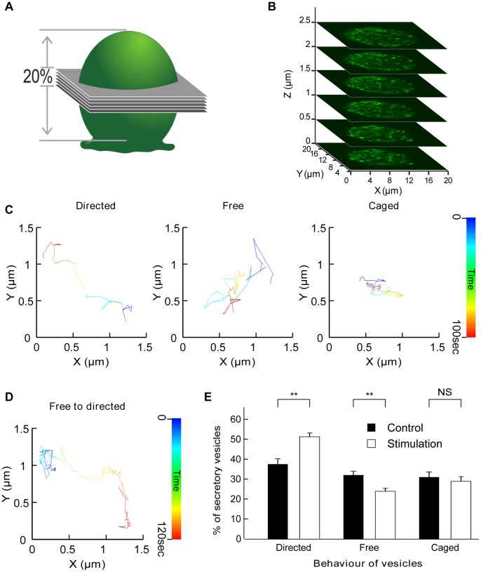
How neurosecretory cells spatially adjust their secretory vesicle pools to replenish those that have fused and released their hormonal content is currently unknown. Here we designed a novel set of image analyses to map the probability of tracked organelles undergoing a specific type of movement (free, caged or directed). We then applied our analysis to time-lapse z-stack confocal imaging of secretory vesicles from bovine Chromaffin cells to map the global changes in vesicle motion and directionality occurring upon secretagogue stimulation. We report a defined region abutting the cortical actin network that actively transports secretory vesicles and is dissipated by actin and microtubule depolymerizing drugs. The directionality of this "conveyor belt" towards the cell surface is activated by stimulation. Actin and microtubule networks therefore cooperatively probe the microenvironment to transport secretory vesicles to the periphery, providing a mechanism whereby cells globally adjust their vesicle pools in response to secretagogue stimulation.
神经分泌细胞如何在空间上调整其分泌小泡池,以补充已经融合并释放其激素内容物的小泡池,目前尚不清楚。在这里,我们设计了一套新的图像分析方法来绘制被跟踪细胞器经历特定类型运动(自由、束缚或定向)的概率。然后,我们将我们的分析应用于牛嗜铬细胞分泌小泡的延时 z -stack 共聚焦成像,以绘制在激动剂刺激下发生的囊泡运动和方向性的全局变化。我们报告了一个紧邻皮质肌动蛋白网络的定义区域,该区域主动运输分泌小泡,并被肌动蛋白和微管解聚药物消耗。这个“输送带”朝着细胞表面的方向性是由刺激激活的。因此,肌动蛋白和微管网络合作探测微环境,将分泌小泡运输到外围,为细胞提供了一种机制,使它们能够根据激动剂刺激全局调整其囊泡池。