Fouda Abdelrahman Y, Kozak Anna, Alhusban Ahmed, Switzer Jeffrey A, Fagan Susan C
Charlie Norwood VA Medical Center , College of Pharmacy, University of Georgia and Center for Pharmacy and Experimental Therapeutics, Augusta, GA, USA.
Exp Transl Stroke Med. 2013 Nov 13;5(1):12. doi: 10.1186/2040-7378-5-12.
Exogenous administration of the anti-inflammatory cytokine, interleukin 10 (IL-10), is known to promote neuroprotection and mitigate neuroinflammation after ischemia. However, endogenous expression and localization of IL-10 and its receptor (IL-10R) in the post-ischemic brain are still to be elucidated. In this investigation we aimed at determining the temporospatial expression of IL-10 in the rat brain relative to its systemic levels after ischemic stroke.
Wistar rats were subjected to either permanent (pMCAO) or 3-h temporary (tMCAO) middle cerebral artery occlusion and euthanized at either 24 or 72 h. IL-10/IL-10R levels were quantified in ischemic and contralesional hemispheres and compared to shams using multiplex bead array and Western blotting, respectively. Localization of IL-10/IL-10R with markers for neurons, microglia, astrocytes & endothelial cells were examined using double labeling immunofluorescence. IL-10 was also quantified in the brain tissue of spontaneously hypertensive rats (SHRs) at 24 h after tMCAO.
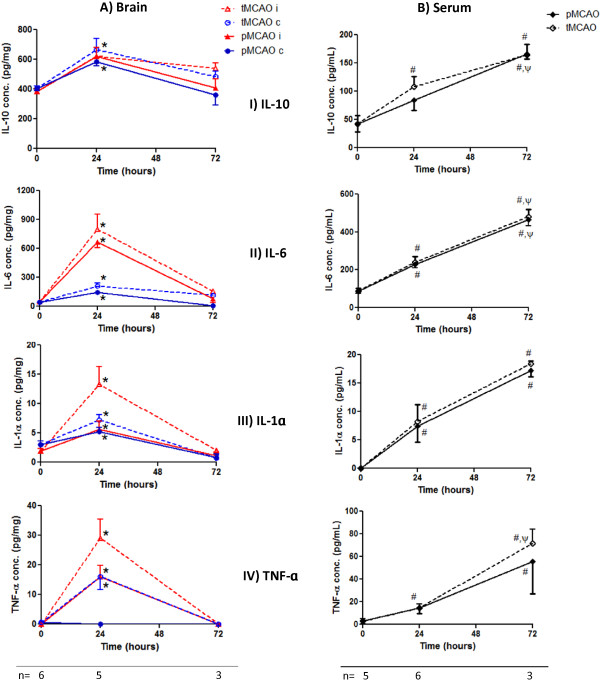
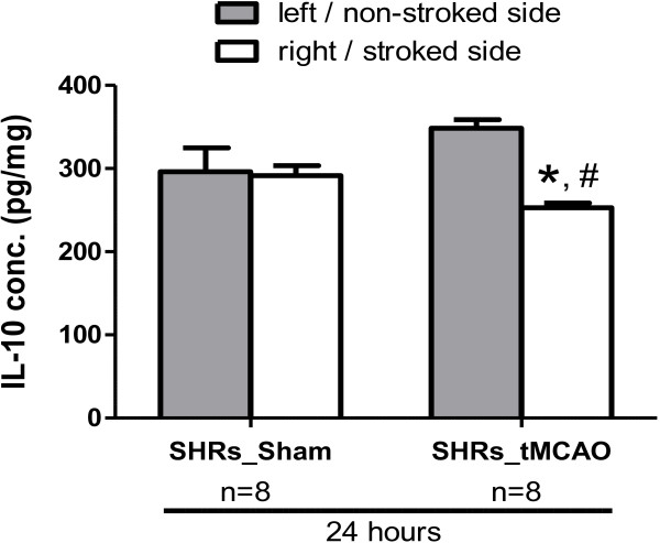
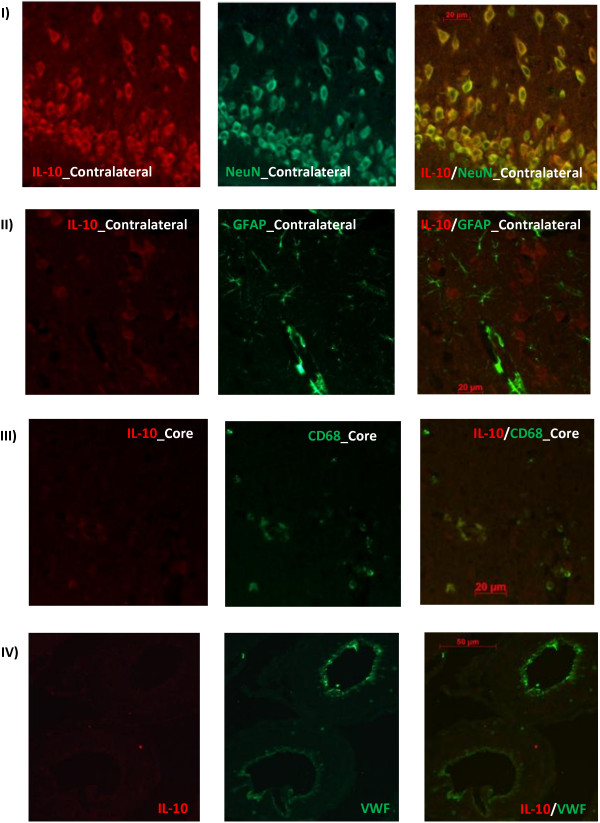
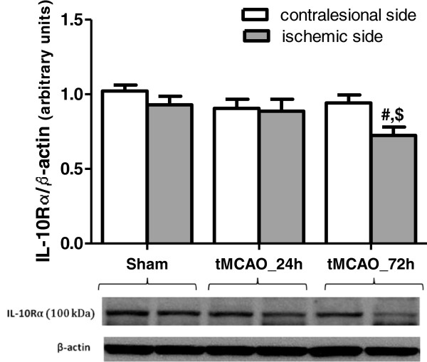
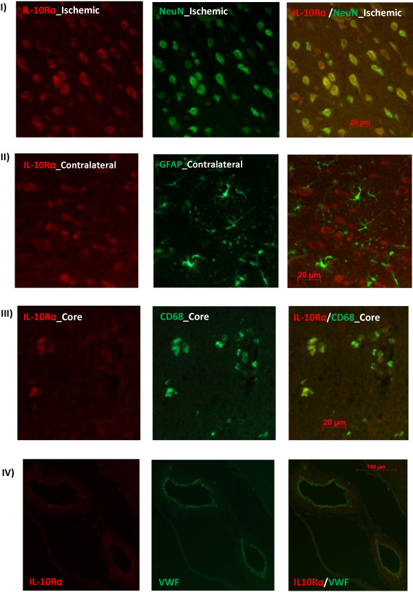
After both pMCAO and tMCAO in Wistars, IL-10 was significantly upregulated in both hemispheres by ≈ 50% at 24 h while IL-10R expression was significantly decreased only at 72 h in the ischemic hemisphere. IL-10 and IL-10R expression highly co-localized with viable neurons in the ischemic penumbra and contralesional hemisphere. In hypertensive rats, IL-10 showed no significant contralesional upregulation and declined significantly in the ischemic side at 24 h post-ischemia.
Our data highlights the involvement of the ischemic and contralesional neurons in the endogenous anti-inflammatory response after ischemic stroke through increased production of IL-10. This increase in IL-10 is blunted in hypertensive animals and may contribute to worse outcomes.
已知外源性给予抗炎细胞因子白细胞介素10(IL-10)可促进神经保护并减轻缺血后的神经炎症。然而,缺血后脑内IL-10及其受体(IL-10R)的内源性表达和定位仍有待阐明。在本研究中,我们旨在确定缺血性卒中后大鼠脑内IL-10相对于其全身水平的时空表达。
将Wistar大鼠进行永久性(pMCAO)或3小时暂时性(tMCAO)大脑中动脉闭塞,并在24小时或72小时处安乐死。分别使用多重珠阵列和蛋白质印迹法对缺血半球和对侧半球的IL-10/IL-10R水平进行定量,并与假手术组进行比较。使用双标免疫荧光检查IL-10/IL-10R与神经元、小胶质细胞、星形胶质细胞和内皮细胞标志物的定位。还对tMCAO后24小时的自发性高血压大鼠(SHR)脑组织中的IL-10进行了定量。
在Wistar大鼠中,pMCAO和tMCAO后,两个半球的IL-10在24小时均显著上调约50%,而IL-10R表达仅在缺血半球的72小时显著降低。IL-10和IL-10R表达在缺血半暗带和对侧半球中与存活神经元高度共定位。在高血压大鼠中,IL-10在对侧没有显著上调,并且在缺血后24小时缺血侧显著下降。
我们的数据表明,缺血性卒中和对侧神经元通过增加IL-10的产生参与内源性抗炎反应。高血压动物中IL-10的这种增加减弱,可能导致更差的结果。