• 文献检索
  • 文档翻译
  • 深度研究
  • 学术资讯
  • Suppr Zotero 插件Zotero 插件
  • 邀请有礼
  • 套餐&价格
  • 历史记录
应用&插件
Suppr Zotero 插件Zotero 插件浏览器插件Mac 客户端Windows 客户端微信小程序
定价
高级版会员购买积分包购买API积分包
服务
文献检索文档翻译深度研究API 文档MCP 服务
关于我们
关于 Suppr公司介绍联系我们用户协议隐私条款
关注我们

Suppr 超能文献

核心技术专利:CN118964589B侵权必究
粤ICP备2023148730 号-1Suppr @ 2026

文献检索

告别复杂PubMed语法,用中文像聊天一样搜索,搜遍4000万医学文献。AI智能推荐,让科研检索更轻松。

立即免费搜索

文件翻译

保留排版,准确专业,支持PDF/Word/PPT等文件格式,支持 12+语言互译。

免费翻译文档

深度研究

AI帮你快速写综述,25分钟生成高质量综述,智能提取关键信息,辅助科研写作。

立即免费体验

原发性眼内淋巴瘤患者眼部的频域光学相干断层扫描及眼底自发荧光检查结果

Spectral-domain optical coherence tomographic and fundus autofluorescence findings in eyes with primary intraocular lymphoma.

作者信息

Egawa Mariko, Mitamura Yoshinori, Hayashi Yuki, Naito Takeshi

机构信息

Department of Ophthalmology, Institute of Health Biosciences, The University of Tokushima Graduate School, Tokushima, Japan.

出版信息

Clin Ophthalmol. 2014 Jan 31;8:335-41. doi: 10.2147/OPTH.S58114. eCollection 2014.

DOI:10.2147/OPTH.S58114
PMID:24520190
原文链接:https://pmc.ncbi.nlm.nih.gov/articles/PMC3917953/
Abstract

BACKGROUND

The purpose of this study was to evaluate the findings on spectral-domain optical coherence tomography (SD-OCT) and fundus autofluorescence (FAF) in three eyes with primary intraocular lymphoma (PIOL).

METHODS

The medical records of three eyes from three patients with biopsy-proven PIOL and retinal infiltrations were reviewed. The SD-OCT and fluorescein angiographic findings were evaluated in the three eyes and FAF images in two eyes.

RESULTS

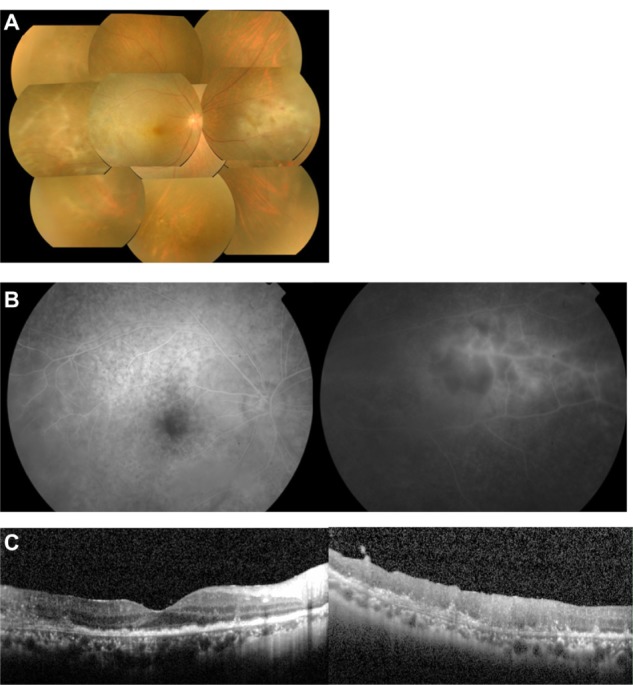
The PIOL in the three patients was monocular. Vitreous opacities and retinal infiltrations were observed in the three eyes, and iritis was present in two eyes. The cytologic diagnosis was class V in two eyes and class III in one eye. The interleukin-10/interleukin-6 ratio was >1.0 in the vitreous and aqueous humor of the three eyes. The FAF images for two eyes showed abnormal granular hyperautofluorescence and hypoautofluorescence which were the reverse of the pattern in the fluorescein angiographic images. In all three eyes, SD-OCT showed hyper-reflective infiltrations at the level of the retinal pigment epithelium (RPE), a separation of the Bruch membrane from the RPE, damage to the RPE, disruption of the photoreceptor inner segment/outer segment junction, and multiple hyper-reflective signals in the inner retina.

CONCLUSION

Because of the characteristic FAF and SD-OCT findings in these eyes with PIOL, we suggest that these noninvasive methods may be used for a rapid diagnosis of PIOL and also for understanding the pathology of PIOL.

摘要

背景

本研究旨在评估原发性眼内淋巴瘤(PIOL)患者三只眼中的频域光学相干断层扫描(SD-OCT)和眼底自发荧光(FAF)检查结果。

方法

回顾了三名经活检证实患有PIOL且伴有视网膜浸润的患者的三只眼的病历。对这三只眼的SD-OCT和荧光素血管造影检查结果进行了评估,并对其中两只眼进行了FAF图像分析。

结果

三名患者的PIOL均为单眼发病。三只眼中均观察到玻璃体混浊和视网膜浸润,两只眼中存在虹膜炎。细胞学诊断为两只眼为V级,一只眼为III级。三只眼的玻璃体和房水中白细胞介素-10/白细胞介素-6比值均>1.0。两只眼的FAF图像显示异常的颗粒状高自发荧光和低自发荧光,与荧光素血管造影图像的表现相反。在所有三只眼中,SD-OCT均显示视网膜色素上皮(RPE)层有高反射性浸润、Bruch膜与RPE分离、RPE损伤、光感受器内段/外段连接破坏以及视网膜内层有多个高反射信号。

结论

鉴于这些PIOL患眼中FAF和SD-OCT的特征性表现,我们认为这些非侵入性方法可用于PIOL的快速诊断以及了解PIOL的病理情况。

https://cdn.ncbi.nlm.nih.gov/pmc/blobs/6926/3917953/54e351d9faad/opth-8-335Fig1.jpg

相似文献

1
Spectral-domain optical coherence tomographic and fundus autofluorescence findings in eyes with primary intraocular lymphoma.原发性眼内淋巴瘤患者眼部的频域光学相干断层扫描及眼底自发荧光检查结果
Clin Ophthalmol. 2014 Jan 31;8:335-41. doi: 10.2147/OPTH.S58114. eCollection 2014.
2
Changes of fundus autofluorescence and spectral-domain optical coherence tomographic findings after treatment of primary intraocular lymphoma.原发性眼内淋巴瘤治疗后眼底自发荧光及频域光学相干断层扫描结果的变化
J Ophthalmic Inflamm Infect. 2014 Feb 22;4(1):7. doi: 10.1186/1869-5760-4-7.
3
Fundus autofluorescence patterns in primary intraocular lymphoma.原发性眼内淋巴瘤的眼底自发荧光模式。
Retina. 2014 Feb;34(2):366-72. doi: 10.1097/IAE.0b013e31829977fa.
4
Acute central serous chorioretinopathy: a correlation study between fundus autofluorescence and spectral-domain OCT.急性中心性浆液性脉络膜视网膜病变:眼底自发荧光与光谱域光学相干断层扫描的相关性研究
Graefes Arch Clin Exp Ophthalmol. 2015 Nov;253(11):1889-97. doi: 10.1007/s00417-014-2899-5. Epub 2015 Jan 8.
5
Spectral domain optical coherence tomography and fundus autofluorescence findings in cytomegalovirus retinitis in HIV-infected patients.HIV感染患者巨细胞病毒性视网膜炎的频域光学相干断层扫描和眼底自发荧光检查结果
Jpn J Ophthalmol. 2018 May;62(3):373-389. doi: 10.1007/s10384-018-0574-9. Epub 2018 Feb 14.
6
Retinal pigment epithelial changes in chronic Vogt-Koyanagi-Harada disease: fundus autofluorescence and spectral domain-optical coherence tomography findings.慢性 Vogt-小柳原田病的视网膜色素上皮变化:眼底自发荧光和谱域光学相干断层扫描的发现。
Retina. 2010 Jan;30(1):33-41. doi: 10.1097/IAE.0b013e3181c5970d.
7
Diabetic retinal pigment epitheliopathy: fundus autofluorescence and spectral-domain optical coherence tomography findings.糖尿病性视网膜色素上皮病变:眼底自发荧光及光谱域光学相干断层扫描结果
Graefes Arch Clin Exp Ophthalmol. 2016 Oct;254(10):1931-1940. doi: 10.1007/s00417-016-3336-8. Epub 2016 Apr 6.
8
Fundus autofluorescence patterns in eyes with primary intraocular lymphoma.原发性眼内淋巴瘤眼的眼底自发荧光模式。
Retina. 2010 Jan;30(1):23-32. doi: 10.1097/IAE.0b013e3181b408a2.
9
Characteristic optical coherence tomography findings in patients with primary vitreoretinal lymphoma: a novel aid to early diagnosis.原发性玻璃体视网膜淋巴瘤患者的特征性光学相干断层扫描表现:一种有助于早期诊断的新方法。
Br J Ophthalmol. 2018 Oct;102(10):1362-1366. doi: 10.1136/bjophthalmol-2017-311612. Epub 2018 Jan 6.
10
A systematic comparison of spectral-domain optical coherence tomography and fundus autofluorescence in patients with geographic atrophy.光谱域光学相干断层扫描与眼底自发荧光在地图样萎缩患者中的系统比较。
Ophthalmology. 2011 Sep;118(9):1844-51. doi: 10.1016/j.ophtha.2011.01.043. Epub 2011 Apr 15.

引用本文的文献

1
Primary vitreoretinal lymphoma: diagnosis, treatment, and prognosis-a review of current knowledge and future directions.原发性玻璃体视网膜淋巴瘤:诊断、治疗及预后——当前知识与未来方向综述
Blood Sci. 2025 May 1;7(2):e00233. doi: 10.1097/BS9.0000000000000233. eCollection 2025 Jun.
2
Clinical Features and Possible Origin of Preretinal Deposits in Different Ocular Diseases and Events: A Narrative Review.不同眼部疾病和事件中视网膜前沉积物的临床特征及可能来源:一项叙述性综述
Ophthalmol Ther. 2023 Apr;12(2):737-753. doi: 10.1007/s40123-023-00674-4. Epub 2023 Feb 16.
3
Multimodal diagnostic imaging in primary vitreoretinal lymphoma.

本文引用的文献

1
Longitudinal spectral domain optical coherence tomography changes in eyes with intraocular lymphoma.眼内淋巴瘤患者眼部的纵向光谱域光学相干断层扫描变化
J Ophthalmic Inflamm Infect. 2013 Sep 8;3(1):59. doi: 10.1186/1869-5760-3-59.
2
Fundus autofluorescence patterns in primary intraocular lymphoma.原发性眼内淋巴瘤的眼底自发荧光模式。
Retina. 2014 Feb;34(2):366-72. doi: 10.1097/IAE.0b013e31829977fa.
3
Clinical features and diagnostic significance of the intraocular fluid of 217 patients with intraocular lymphoma.217 例眼内淋巴瘤患者眼内液的临床特征和诊断意义。
原发性玻璃体视网膜淋巴瘤的多模态诊断成像
Int J Retina Vitreous. 2022 Aug 26;8(1):58. doi: 10.1186/s40942-022-00405-0.
4
Optical Coherence Tomography Benefits the Diagnosis and Follow-Up of Primary Central Nervous System Lymphoma with Intraocular Involvement.光学相干断层扫描有助于原发性中枢神经系统淋巴瘤合并眼内受累的诊断及随访。
Cancer Manag Res. 2022 Mar 5;14:1007-1018. doi: 10.2147/CMAR.S353142. eCollection 2022.
5
Clinical Features, Diagnosis, Management and Prognosis of Primary Intraocular Lymphoma.原发性眼内淋巴瘤的临床特征、诊断、治疗与预后
Front Oncol. 2022 Feb 3;12:808511. doi: 10.3389/fonc.2022.808511. eCollection 2022.
6
Multimodal imaging of secondary vitreoretinal lymphoma with optic neuritis and retinal vasculitis.伴有视神经炎和视网膜血管炎的继发性玻璃体视网膜淋巴瘤的多模态成像
Am J Ophthalmol Case Rep. 2020 Apr 8;18:100696. doi: 10.1016/j.ajoc.2020.100696. eCollection 2020 Jun.
7
Primary vitreoretinal lymphoma.原发性玻璃体视网膜淋巴瘤
Saudi J Ophthalmol. 2019 Jan-Mar;33(1):66-80. doi: 10.1016/j.sjopt.2018.12.008. Epub 2019 Jan 14.
8
Primary vitreoretinal lymphoma: prevalence, impact, and management challenges.原发性玻璃体视网膜淋巴瘤:患病率、影响及管理挑战
Clin Ophthalmol. 2019 Feb 14;13:353-364. doi: 10.2147/OPTH.S159014. eCollection 2019.
9
A Case of Primary Vitreous Retinal Lymphoma.原发性玻璃体视网膜淋巴瘤1例。
Chin Med J (Engl). 2018 Jan 5;131(1):113-114. doi: 10.4103/0366-6999.221281.
10
Recent progress in the diagnosis and treatment of primary vitreoretinal lymphoma.原发性玻璃体视网膜淋巴瘤诊断与治疗的最新进展
Taiwan J Ophthalmol. 2016 Oct-Dec;6(4):170-176. doi: 10.1016/j.tjo.2016.05.002. Epub 2016 Jun 27.
Jpn J Ophthalmol. 2012 Jul;56(4):383-9. doi: 10.1007/s10384-012-0150-7. Epub 2012 Jun 5.
4
Spatial correlation between hyperpigmentary changes on color fundus photography and hyperreflective foci on SDOCT in intermediate AMD.中度年龄相关性黄斑变性患者彩色眼底照片上的色素沉着改变与光谱域光学相干断层扫描上的高反射灶之间的空间相关性。
Invest Ophthalmol Vis Sci. 2012 Jul 9;53(8):4626-33. doi: 10.1167/iovs.12-9813.
5
Retinal optical coherence tomography manifestations of intraocular lymphoma.眼内淋巴瘤的视网膜光学相干断层扫描表现
J Ophthalmic Inflamm Infect. 2012 Dec;2(4):215-8. doi: 10.1007/s12348-012-0072-z. Epub 2012 Apr 4.
6
Diagnostic imaging in patients with retinitis pigmentosa.视网膜色素变性患者的诊断性影像学检查
J Med Invest. 2012;59(1-2):1-11. doi: 10.2152/jmi.59.1.
7
Subretinal lymphomatous infiltration in primary intraocular lymphoma revealed by optical coherence tomography.光学相干断层扫描显示原发性眼内淋巴瘤的视网膜下淋巴瘤浸润。
Graefes Arch Clin Exp Ophthalmol. 2011 Sep;249(9):1425-7. doi: 10.1007/s00417-011-1669-x. Epub 2011 Apr 21.
8
Endosopic autofluorescence imaging is useful for the differential diagnosis of intestinal lymphomas resembling lymphoid hyperplasia.内镜自体荧光成像是一种有用的方法,可用于鉴别类似于淋巴组织增生的肠道淋巴瘤。
J Clin Gastroenterol. 2011 Jul;45(6):507-13. doi: 10.1097/MCG.0b013e3181fbe22a.
9
Optical coherence tomographic evaluation of foveal hard exudates in patients with diabetic maculopathy accompanying macular detachment.光学相干断层扫描评价伴有黄斑脱离的糖尿病性黄斑病变患者的中心凹硬性渗出物。
Ophthalmology. 2010 Oct;117(10):1996-2002. doi: 10.1016/j.ophtha.2010.06.019. Epub 2010 Aug 17.
10
Changes of fundus autofluorescence, photoreceptor inner and outer segment junction line, and visual function in patients with retinitis pigmentosa.色素性视网膜炎患者的眼底自发荧光、光感受器内外节连接带和视觉功能的变化。
Clin Exp Ophthalmol. 2010 Aug;38(6):597-604. doi: 10.1111/j.1442-9071.2010.02321.x. Epub 2010 Apr 29.