Donati Abele, Damiani Elisa, Luchetti Michele, Domizi Roberta, Scorcella Claudia, Carsetti Andrea, Gabbanelli Vincenzo, Carletti Paola, Bencivenga Rosella, Vink Hans, Adrario Erica, Piagnerelli Michael, Gabrielli Armando, Pelaia Paolo, Ince Can
Crit Care. 2014 Feb 17;18(1):R33. doi: 10.1186/cc13730.
Microvascular alterations impair tissue oxygenation during sepsis. A red blood cell (RBC) transfusion increases oxygen (O2) delivery but rarely improves tissue O2 uptake in patients with sepsis. Possible causes include RBC alterations due to prolonged storage or residual leukocyte-derived inflammatory mediators. The aim of this study was to compare the effects of two types of transfused RBCs on microcirculation in patients with sepsis.
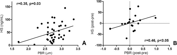
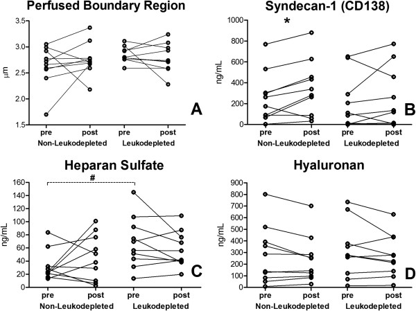
In a prospective randomized trial, 20 patients with sepsis were divided into two separate groups and received either non-leukodepleted (n = 10) or leukodepleted (n = 10) RBC transfusions. Microvascular density and perfusion were assessed with sidestream dark field (SDF) imaging sublingually, before and 1 hour after transfusions. Thenar tissue O2 saturation (StO2) and tissue hemoglobin index (THI) were determined with near-infrared spectroscopy, and a vascular occlusion test was performed. The microcirculatory perfused boundary region was assessed in SDF images as an index of glycocalyx damage, and glycocalyx compounds (syndecan-1, hyaluronan, and heparan sulfate) were measured in the serum.
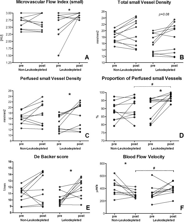
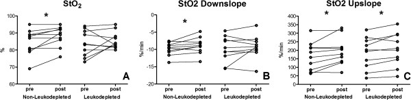
No differences were observed in microvascular parameters at baseline and after transfusion between the groups, except for the proportion of perfused vessels (PPV) and blood flow velocity, which were higher after transfusion in the leukodepleted group. Microvascular flow index in small vessels (MFI) and blood flow velocity exhibited different responses to transfusion between the two groups (P = 0.03 and P = 0.04, respectively), with a positive effect of leukodepleted RBCs. When within-group changes were examined, microcirculatory improvement was observed only in patients who received leukodepleted RBC transfusion as suggested by the increase in De Backer score (P = 0.02), perfused vessel density (P = 0.04), PPV (P = 0.01), and MFI (P = 0.04). Blood flow velocity decreased in the non-leukodepleted group (P = 0.03). THI and StO2 upslope increased in both groups. StO2 and StO2 downslope increased in patients who received non-leukodepleted RBC transfusions. Syndecan-1 increased after the transfusion of non-leukodepleted RBCs (P = 0.03).
This study does not show a clear superiority of leukodepleted over non-leukodepleted RBC transfusions on microvascular perfusion in patients with sepsis, although it suggests a more favorable effect of leukodepleted RBCs on microcirculatory convective flow. Further studies are needed to confirm these findings.
ClinicalTrials.gov, NCT01584999.
脓毒症期间微血管改变会损害组织氧合。红细胞(RBC)输血可增加氧(O₂)输送,但在脓毒症患者中很少能改善组织O₂摄取。可能的原因包括由于长期储存导致的RBC改变或残留的白细胞衍生炎症介质。本研究的目的是比较两种类型的输注RBC对脓毒症患者微循环的影响。
在一项前瞻性随机试验中,20例脓毒症患者被分为两个独立的组,分别接受非去白细胞(n = 10)或去白细胞(n = 10)RBC输血。在输血前和输血后1小时,使用侧流暗视野(SDF)成像舌下评估微血管密度和灌注。用近红外光谱法测定鱼际组织氧饱和度(StO₂)和组织血红蛋白指数(THI),并进行血管闭塞试验。在SDF图像中评估微循环灌注边界区域作为糖萼损伤的指标,并测定血清中的糖萼化合物(syndecan - 1、透明质酸和硫酸乙酰肝素)。
两组在基线和输血后的微血管参数上未观察到差异,但去白细胞组输血后灌注血管比例(PPV)和血流速度较高。两组小血管的微血管血流指数(MFI)和血流速度对输血表现出不同的反应(分别为P = 0.03和P = 0.04),去白细胞RBC有积极作用。当检查组内变化时,如De Backer评分增加(P = 0.02)、灌注血管密度增加(P = 0.04)、PPV增加(P = 0.01)和MFI增加(P = 0.04)所示,仅接受去白细胞RBC输血的患者观察到微循环改善。非去白细胞组血流速度下降(P = 0.03)。两组的THI和StO₂上升斜率均增加。接受非去白细胞RBC输血的患者StO₂和StO₂下降斜率增加。非去白细胞RBC输血后syndecan - 1增加(P = 0.03)。
本研究未显示去白细胞RBC输血在脓毒症患者微血管灌注方面明显优于非去白细胞RBC输血,尽管提示去白细胞RBC对微循环对流有更有利的影响。需要进一步研究来证实这些发现。
ClinicalTrials.gov,NCT01584999。