• 文献检索
  • 文档翻译
  • 深度研究
  • 学术资讯
  • Suppr Zotero 插件Zotero 插件
  • 邀请有礼
  • 套餐&价格
  • 历史记录
应用&插件
Suppr Zotero 插件Zotero 插件浏览器插件Mac 客户端Windows 客户端微信小程序
定价
高级版会员购买积分包购买API积分包
服务
文献检索文档翻译深度研究API 文档MCP 服务
关于我们
关于 Suppr公司介绍联系我们用户协议隐私条款
关注我们

Suppr 超能文献

核心技术专利:CN118964589B侵权必究
粤ICP备2023148730 号-1Suppr @ 2026

文献检索

告别复杂PubMed语法,用中文像聊天一样搜索,搜遍4000万医学文献。AI智能推荐,让科研检索更轻松。

立即免费搜索

文件翻译

保留排版,准确专业,支持PDF/Word/PPT等文件格式,支持 12+语言互译。

免费翻译文档

深度研究

AI帮你快速写综述,25分钟生成高质量综述,智能提取关键信息,辅助科研写作。

立即免费体验

量化心肌细胞肥大差异调节的表型筛选确定了CITED4对心肌细胞伸长的调节作用。

Phenotypic screen quantifying differential regulation of cardiac myocyte hypertrophy identifies CITED4 regulation of myocyte elongation.

作者信息

Ryall Karen A, Bezzerides Vassilios J, Rosenzweig Anthony, Saucerman Jeffrey J

机构信息

Department of Biomedical Engineering, University of Virginia, VA, USA.

Department of Cardiology, Children's Hospital Boston, Harvard Medical School, Boston, MA, USA.

出版信息

J Mol Cell Cardiol. 2014 Jul;72:74-84. doi: 10.1016/j.yjmcc.2014.02.013. Epub 2014 Mar 5.

DOI:10.1016/j.yjmcc.2014.02.013
PMID:24613264
原文链接:https://pmc.ncbi.nlm.nih.gov/articles/PMC4078663/
Abstract

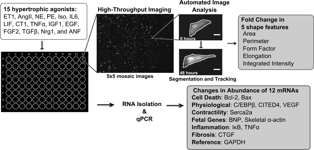
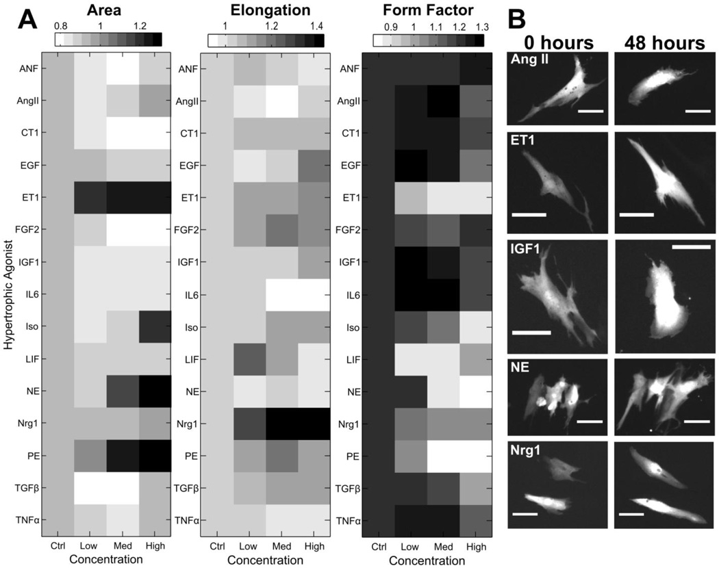
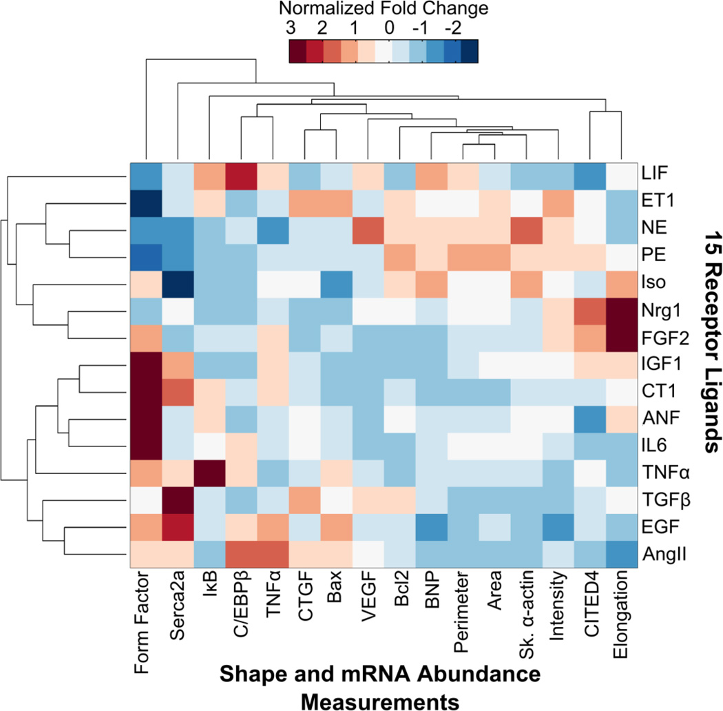
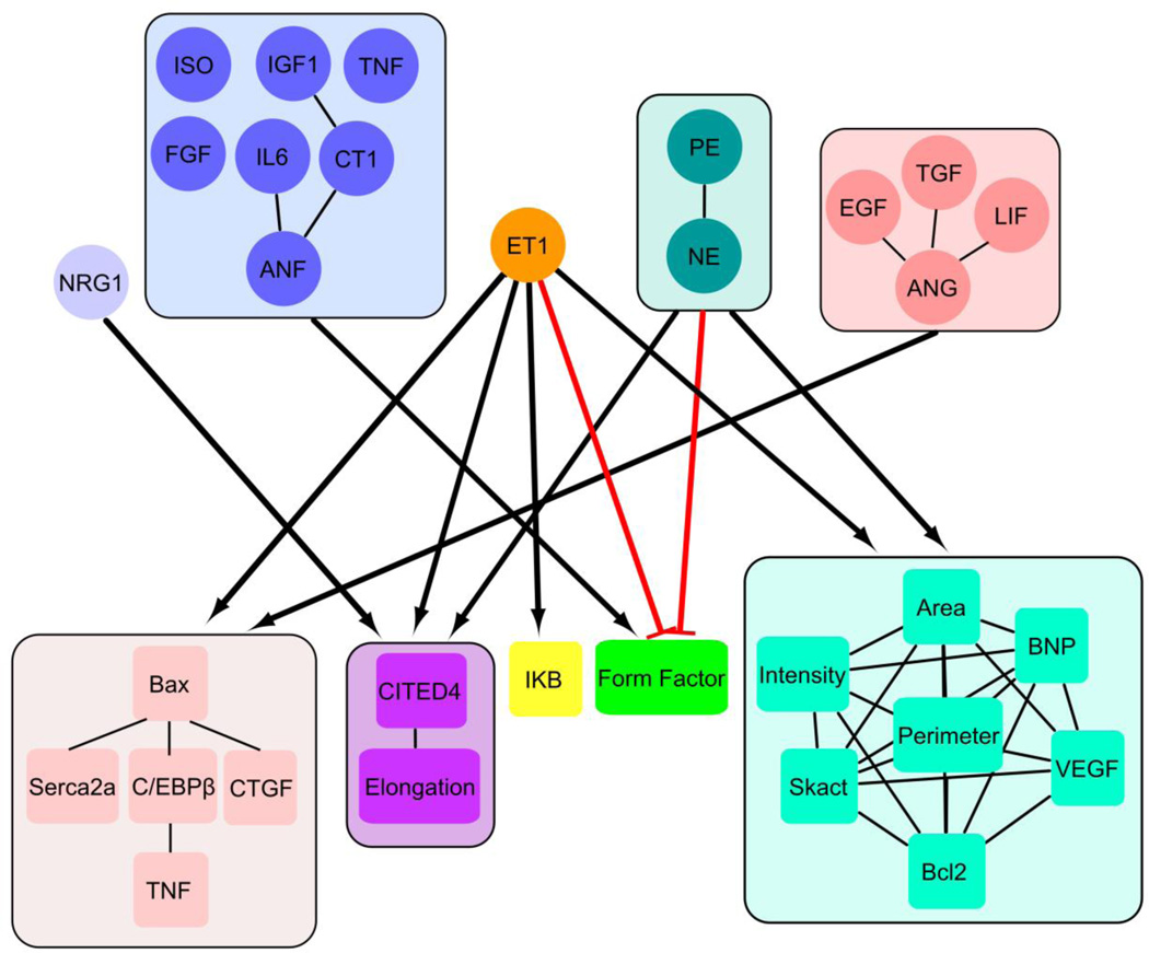
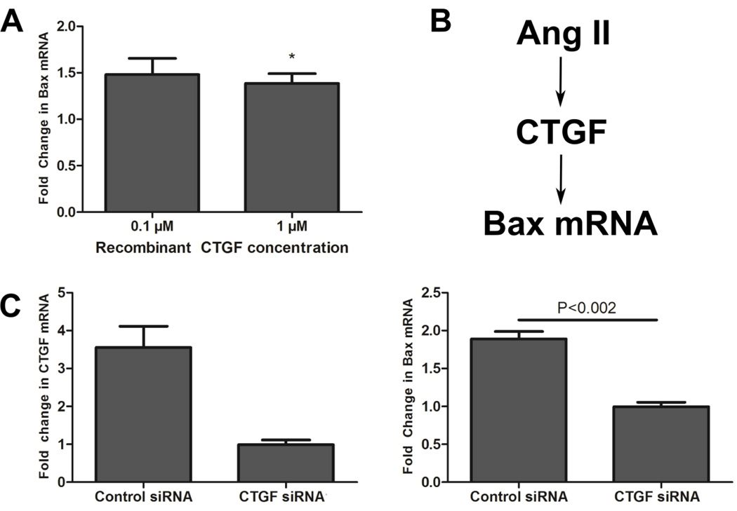
Cardiac hypertrophy is controlled by a highly connected signaling network with many effectors of cardiac myocyte size. Quantification of the contribution of individual pathways to specific changes in shape and transcript abundance is needed to better understand hypertrophy signaling and to improve heart failure therapies. We stimulated cardiac myocytes with 15 hypertrophic agonists and quantitatively characterized differential regulation of 5 shape features using high-throughput microscopy and transcript levels of 12 genes using qPCR. Transcripts measured were associated with phenotypes including fibrosis, cell death, contractility, proliferation, angiogenesis, inflammation, and the fetal cardiac gene program. While hypertrophy pathways are highly connected, the agonist screen revealed distinct hypertrophy phenotypic signatures for the 15 receptor agonists. We then used k-means clustering of inputs and outputs to identify a network map linking input modules to output modules. Five modules were identified within inputs and outputs with many maladaptive outputs grouping together in one module: Bax, C/EBPβ, Serca2a, TNFα, and CTGF. Subsequently, we identified mechanisms underlying two correlations revealed in the agonist screen: correlation between regulators of fibrosis and cell death signaling (CTGF and Bax mRNA) caused by AngII; and myocyte proliferation (CITED4 mRNA) and elongation caused by Nrg1. Follow-up experiments revealed positive regulation of Bax mRNA level by CTGF and an incoherent feedforward loop linking Nrg1, CITED4 and elongation. With this agonist screen, we identified the most influential inputs in the cardiac hypertrophy signaling network for a variety of features related to pathological and protective hypertrophy signaling and shared regulation among cardiac myocyte phenotypes.

摘要

心脏肥大受一个高度连接的信号网络控制,该网络中有许多影响心肌细胞大小的效应器。为了更好地理解肥大信号并改善心力衰竭治疗,需要量化各个信号通路对形状和转录本丰度特定变化的贡献。我们用15种肥大激动剂刺激心肌细胞,使用高通量显微镜对5种形状特征的差异调节进行定量表征,并使用qPCR对12个基因的转录水平进行定量表征。所测量的转录本与包括纤维化、细胞死亡、收缩性、增殖、血管生成、炎症和胎儿心脏基因程序等表型相关。虽然肥大信号通路高度连接,但激动剂筛选揭示了15种受体激动剂不同的肥大表型特征。然后,我们使用输入和输出的k均值聚类来识别一个将输入模块与输出模块连接起来的网络图谱。在输入和输出中识别出五个模块,许多适应不良的输出聚集在一个模块中:Bax、C/EBPβ、Serca2a、TNFα和CTGF。随后,我们确定了激动剂筛选中揭示的两种相关性的潜在机制:AngII引起的纤维化调节因子与细胞死亡信号(CTGF和Bax mRNA)之间的相关性;以及Nrg1引起的心肌细胞增殖(CITED4 mRNA)和伸长。后续实验揭示了CTGF对Bax mRNA水平的正向调节以及连接Nrg1、CITED4和伸长的非相干前馈环。通过这种激动剂筛选,我们确定了心脏肥大信号网络中对于与病理性和保护性肥大信号相关的各种特征以及心肌细胞表型之间共享调节最具影响力的输入。

https://cdn.ncbi.nlm.nih.gov/pmc/blobs/2f73/4078663/a3799679ca0b/nihms573718f8.jpg
https://cdn.ncbi.nlm.nih.gov/pmc/blobs/2f73/4078663/574fb6895811/nihms573718f1.jpg
https://cdn.ncbi.nlm.nih.gov/pmc/blobs/2f73/4078663/8e8da15411c7/nihms573718f2.jpg
https://cdn.ncbi.nlm.nih.gov/pmc/blobs/2f73/4078663/4ddafbc40761/nihms573718f3.jpg
https://cdn.ncbi.nlm.nih.gov/pmc/blobs/2f73/4078663/f131fd9c7929/nihms573718f4.jpg
https://cdn.ncbi.nlm.nih.gov/pmc/blobs/2f73/4078663/b1635b2622cb/nihms573718f5.jpg
https://cdn.ncbi.nlm.nih.gov/pmc/blobs/2f73/4078663/40da9426df79/nihms573718f6.jpg
https://cdn.ncbi.nlm.nih.gov/pmc/blobs/2f73/4078663/cd6a7d901fbe/nihms573718f7.jpg
https://cdn.ncbi.nlm.nih.gov/pmc/blobs/2f73/4078663/a3799679ca0b/nihms573718f8.jpg

相似文献

1
Phenotypic screen quantifying differential regulation of cardiac myocyte hypertrophy identifies CITED4 regulation of myocyte elongation.量化心肌细胞肥大差异调节的表型筛选确定了CITED4对心肌细胞伸长的调节作用。
J Mol Cell Cardiol. 2014 Jul;72:74-84. doi: 10.1016/j.yjmcc.2014.02.013. Epub 2014 Mar 5.
2
Automated image analysis identifies signaling pathways regulating distinct signatures of cardiac myocyte hypertrophy.自动化图像分析鉴定出调控心肌细胞肥大不同特征的信号通路。
J Mol Cell Cardiol. 2012 May;52(5):923-30. doi: 10.1016/j.yjmcc.2011.11.009. Epub 2011 Dec 1.
3
Network reconstruction and systems analysis of cardiac myocyte hypertrophy signaling.心肌细胞肥大信号的网络重建和系统分析。
J Biol Chem. 2012 Dec 7;287(50):42259-68. doi: 10.1074/jbc.M112.382937. Epub 2012 Oct 22.
4
Angiotensin II receptors subtypes mediate diverse gene expression profile in adult hypertrophic cardiomyocytes.血管紧张素II受体亚型介导成年肥厚型心肌细胞中不同的基因表达谱。
Clin Exp Pharmacol Physiol. 2007 Nov;34(11):1191-8. doi: 10.1111/j.1440-1681.2007.04694.x.
5
Atorvastatin prevents Angiotensin II induced myocardial hypertrophy in vitro via CCAAT/enhancer-binding protein β.阿托伐他汀通过CCAAT/增强子结合蛋白β在体外预防血管紧张素II诱导的心肌肥大。
Biochem Biophys Res Commun. 2017 Apr 29;486(2):423-430. doi: 10.1016/j.bbrc.2017.03.057. Epub 2017 Mar 15.
6
Inhibition of Apoptosis Signal-Regulating Kinase 1 Attenuates Myocyte Hypertrophy and Fibroblast Collagen Synthesis.凋亡信号调节激酶1的抑制减弱心肌细胞肥大和成纤维细胞胶原蛋白合成。
Heart Lung Circ. 2019 Mar;28(3):495-504. doi: 10.1016/j.hlc.2017.12.001. Epub 2017 Dec 14.
7
Rho-kinase inhibition reverses impaired Ca handling and associated left ventricular dysfunction in pressure overload-induced cardiac hypertrophy.Rho 激酶抑制逆转压力超负荷诱导的心肌肥厚中钙处理受损和相关的左心室功能障碍。
Cell Calcium. 2017 Nov;67:81-90. doi: 10.1016/j.ceca.2017.09.002. Epub 2017 Sep 9.
8
Extracellular signal-regulated kinase activation during cardiac hypertrophy reduces sarcoplasmic/endoplasmic reticulum calcium ATPase 2 (SERCA2) transcription.心脏肥大期间细胞外信号调节激酶的激活会降低肌浆网/内质网钙ATP酶2(SERCA2)的转录。
J Mol Cell Cardiol. 2014 Oct;75:58-63. doi: 10.1016/j.yjmcc.2014.06.018. Epub 2014 Jul 6.
9
eNOS deficient mice develop progressive cardiac hypertrophy with altered cytokine and calcium handling protein expression.内皮型一氧化氮合酶缺乏的小鼠会出现进行性心脏肥大,伴有细胞因子和钙处理蛋白表达改变。
Cardiovasc Toxicol. 2007;7(3):165-77. doi: 10.1007/s12012-007-0028-y.
10
Inducible cAMP early repressor (ICER) is a negative-feedback regulator of cardiac hypertrophy and an important mediator of cardiac myocyte apoptosis in response to beta-adrenergic receptor stimulation.诱导型环磷酸腺苷早期阻遏物(ICER)是心脏肥大的负反馈调节因子,也是响应β-肾上腺素能受体刺激时心肌细胞凋亡的重要介质。
Circ Res. 2003 Jul 11;93(1):12-22. doi: 10.1161/01.RES.0000079794.57578.F1. Epub 2003 Jun 5.

引用本文的文献

1
Identifying metabolic adaptations characteristic of cardiotoxicity using paired transcriptomics and metabolomics data integrated with a computational model of heart metabolism.利用配对转录组学和代谢组学数据并结合心脏代谢计算模型,识别心脏毒性特有的代谢适应性。
PLoS Comput Biol. 2024 Feb 29;20(2):e1011919. doi: 10.1371/journal.pcbi.1011919. eCollection 2024 Feb.
2
Exercise Promotes Tissue Regeneration: Mechanisms Involved and Therapeutic Scope.运动促进组织再生:涉及的机制及治疗范围。
Sports Med Open. 2023 May 6;9(1):27. doi: 10.1186/s40798-023-00573-9.
3
A Kinase Interacting Protein 1 (AKIP1) promotes cardiomyocyte elongation and physiological cardiac remodelling.

本文引用的文献

1
Left ventricular hypertrophy patterns and incidence of heart failure with preserved versus reduced ejection fraction.左心室肥厚模式与射血分数保留与降低的心衰发生率。
Am J Cardiol. 2014 Jan 1;113(1):117-22. doi: 10.1016/j.amjcard.2013.09.028. Epub 2013 Oct 4.
2
Pathological ventricular remodeling: mechanisms: part 1 of 2.病理性心室重构:机制:第 1 部分,共 2 部分。
Circulation. 2013 Jul 23;128(4):388-400. doi: 10.1161/CIRCULATIONAHA.113.001878.
3
NO triggers RGS4 degradation to coordinate angiogenesis and cardiomyocyte growth.
激酶相互作用蛋白 1(AKIP1)促进心肌细胞伸长和生理性心脏重塑。
Sci Rep. 2023 Mar 10;13(1):4046. doi: 10.1038/s41598-023-30514-1.
4
Molecular mechanisms of exercise contributing to tissue regeneration.运动促进组织再生的分子机制。
Signal Transduct Target Ther. 2022 Nov 30;7(1):383. doi: 10.1038/s41392-022-01233-2.
5
Exercise-induced CITED4 expression is necessary for regional remodeling of cardiac microstructural tissue helicity.运动诱导的 CITED4 表达对于心脏微结构组织螺旋的区域性重塑是必需的。
Commun Biol. 2022 Jul 4;5(1):656. doi: 10.1038/s42003-022-03635-y.
6
Exercise: a molecular tool to boost muscle growth and mitochondrial performance in heart failure?运动:促进心力衰竭患者肌肉生长和线粒体功能的分子工具?
Eur J Heart Fail. 2022 Feb;24(2):287-298. doi: 10.1002/ejhf.2407. Epub 2022 Jan 9.
7
C-MORE: A high-content single-cell morphology recognition methodology for liquid biopsies toward personalized cardiovascular medicine.C-MORE:一种用于液体活检的高通量单细胞形态识别方法,旨在实现个体化心血管医学。
Cell Rep Med. 2021 Nov 3;2(11):100436. doi: 10.1016/j.xcrm.2021.100436. eCollection 2021 Nov 16.
8
Integrated Quantitative Phosphoproteomics and Cell-based Functional Screening Reveals Specific Pathological Cardiac Hypertrophy-related Phosphorylation Sites.整合定量磷酸化蛋白质组学和基于细胞的功能筛选揭示特定的病理性心脏肥大相关磷酸化位点。
Mol Cells. 2021 Jul 31;44(7):500-516. doi: 10.14348/molcells.2021.4002.
9
Targets identified from exercised heart: killing multiple birds with one stone.从运动心脏中鉴定出的靶点:一石多鸟。
NPJ Regen Med. 2021 Apr 9;6(1):23. doi: 10.1038/s41536-021-00128-0.
10
Network Analysis Reveals a Distinct Axis of Macrophage Activation in Response to Conflicting Inflammatory Cues.网络分析揭示了在应对冲突性炎症信号时,巨噬细胞激活的一个独特轴。
J Immunol. 2021 Feb 15;206(4):883-891. doi: 10.4049/jimmunol.1901444. Epub 2021 Jan 6.
NO 不会触发 RGS4 降解以协调血管生成和心肌细胞生长。
J Clin Invest. 2013 Apr;123(4):1718-31. doi: 10.1172/JCI65112.
4
Signaling effectors underlying pathologic growth and remodeling of the heart.心脏病理性生长和重构的信号效应子。
J Clin Invest. 2013 Jan;123(1):37-45. doi: 10.1172/JCI62839. Epub 2013 Jan 2.
5
Molecular basis of physiological heart growth: fundamental concepts and new players.生理性心脏生长的分子基础:基本概念和新的参与者。
Nat Rev Mol Cell Biol. 2013 Jan;14(1):38-48. doi: 10.1038/nrm3495.
6
Network reconstruction and systems analysis of cardiac myocyte hypertrophy signaling.心肌细胞肥大信号的网络重建和系统分析。
J Biol Chem. 2012 Dec 7;287(50):42259-68. doi: 10.1074/jbc.M112.382937. Epub 2012 Oct 22.
7
Automated imaging reveals a concentration dependent delay in reversibility of cardiac myocyte hypertrophy.自动化成像显示,心肌细胞肥大的可复性延迟与浓度有关。
J Mol Cell Cardiol. 2012 Aug;53(2):282-90. doi: 10.1016/j.yjmcc.2012.04.016. Epub 2012 May 8.
8
Bax regulates primary necrosis through mitochondrial dynamics.Bax 通过线粒体动力学调节原初坏死。
Proc Natl Acad Sci U S A. 2012 Apr 24;109(17):6566-71. doi: 10.1073/pnas.1201608109. Epub 2012 Apr 9.
9
Une cellule type?一种细胞类型?
J Mol Cell Cardiol. 2012 May;52(5):921-2. doi: 10.1016/j.yjmcc.2012.01.014. Epub 2012 Feb 3.
10
Automated image analysis identifies signaling pathways regulating distinct signatures of cardiac myocyte hypertrophy.自动化图像分析鉴定出调控心肌细胞肥大不同特征的信号通路。
J Mol Cell Cardiol. 2012 May;52(5):923-30. doi: 10.1016/j.yjmcc.2011.11.009. Epub 2011 Dec 1.