Saad Hicham, Gallardo Franck, Dalvai Mathieu, Tanguy-le-Gac Nicolas, Lane David, Bystricky Kerstin
University of Toulouse, UPS, Toulouse, France; Laboratoire de Biologie Moléculaire Eucaryote, CNRS, UMR5099, Toulouse, France.
University of Toulouse, UPS, Toulouse, France; Laboratoire de Biologie Moléculaire Eucaryote, CNRS, UMR5099, Toulouse, France; Institut des Technologies Avancées en sciences du Vivant, ITAV, Toulouse, France.
PLoS Genet. 2014 Mar 13;10(3):e1004187. doi: 10.1371/journal.pgen.1004187. eCollection 2014 Mar.
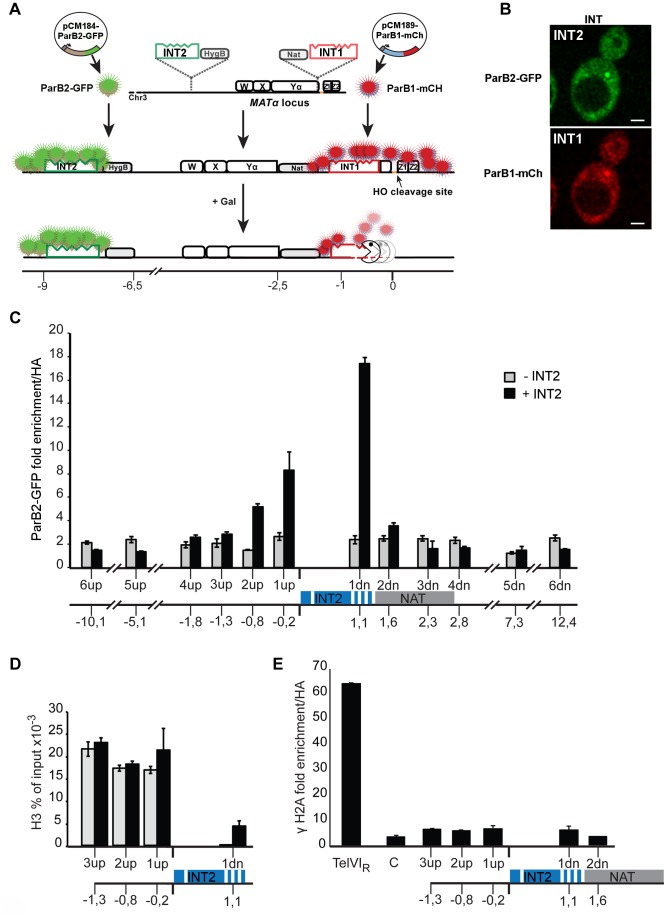
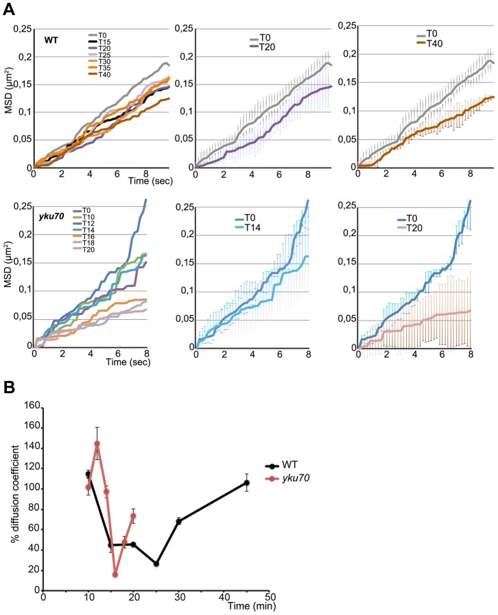
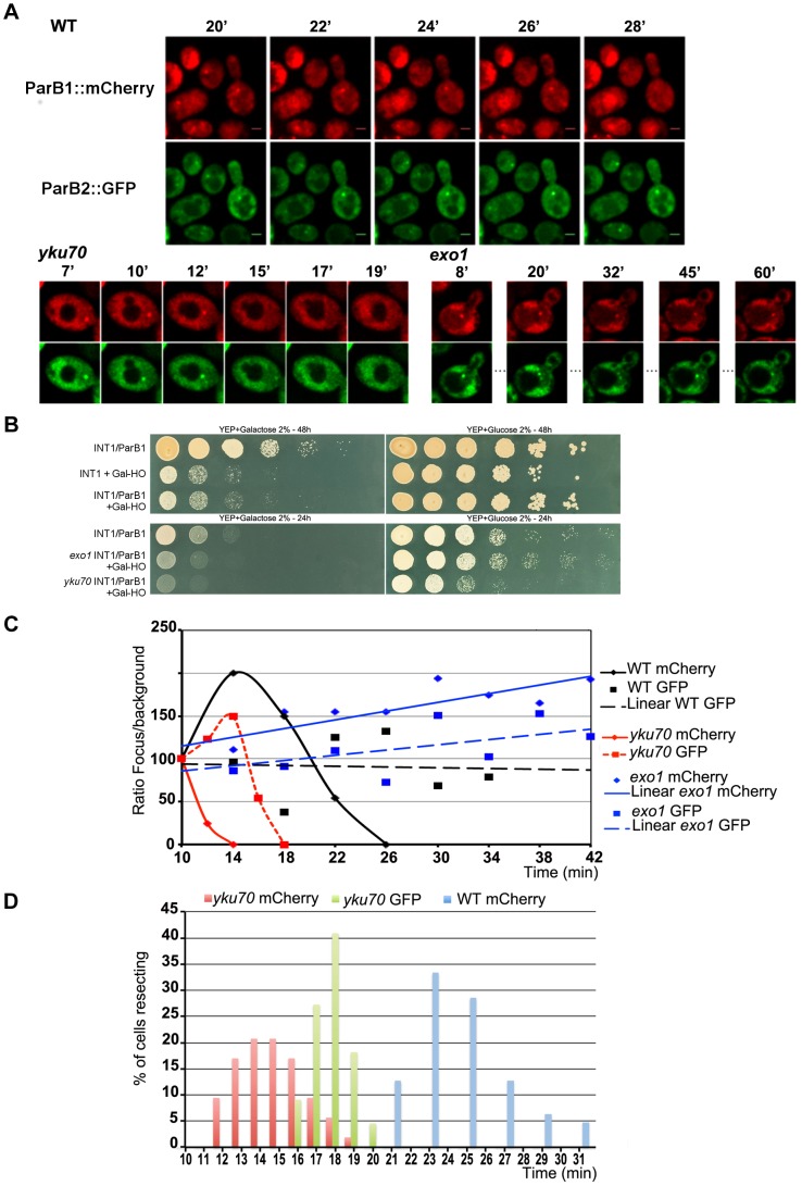
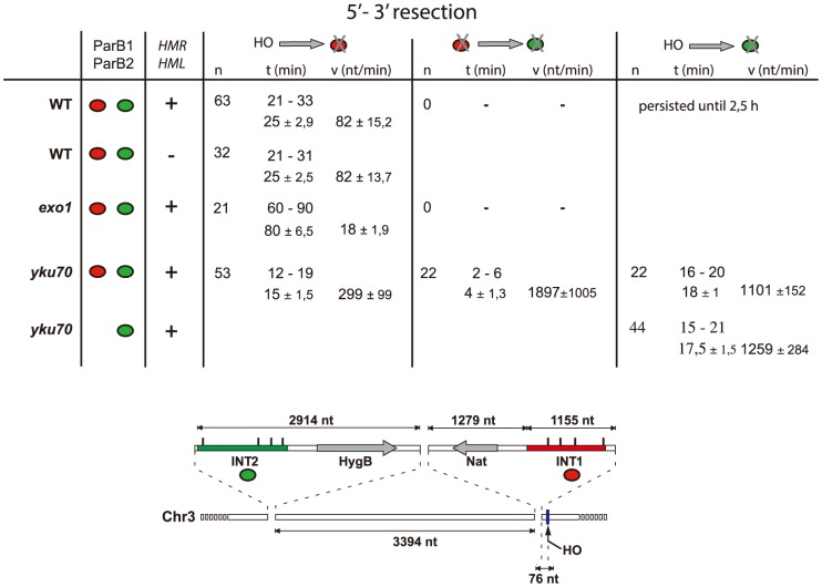
Chromosome breakage is a major threat to genome integrity. The most accurate way to repair DNA double strand breaks (DSB) is homologous recombination (HR) with an intact copy of the broken locus. Mobility of the broken DNA has been seen to increase during the search for a donor copy. Observing chromosome dynamics during the earlier steps of HR, mainly the resection from DSB ends that generates recombinogenic single strands, requires a visualization system that does not interfere with the process, and is small relative to the few kilobases of DNA that undergo processing. Current visualization tools, based on binding of fluorescent repressor proteins to arrays of specific binding sites, have the major drawback that highly-repeated DNA and lengthy stretches of strongly bound protein can obstruct chromatin function. We have developed a new, non-intrusive method which uses protein oligomerization rather than operator multiplicity to form visible foci. By applying it to HO cleavage of the MAT locus on Saccharomyces cerevisiae chromosome III, we provide the first real-time analysis of resection in single living cells. Monitoring the dynamics of a chromatin locus next to a DSB revealed transient confinement of the damaged chromatin region during the very early steps of resection, consistent with the need to keep DNA ends in contact. Resection in a yku70 mutant began ∼ 10 min earlier than in wild type, defining this as the period of commitment to homology-dependent repair. Beyond the insights into the dynamics and mechanism of resection, our new DNA-labelling and -targeting method will be widely applicable to fine-scale analysis of genome organization, dynamics and function in normal and pathological contexts.
染色体断裂是对基因组完整性的重大威胁。修复DNA双链断裂(DSB)最准确的方法是通过同源重组(HR)利用断裂位点的完整拷贝。在寻找供体拷贝的过程中,已观察到断裂DNA的移动性会增加。在HR的早期步骤中观察染色体动态,主要是从DSB末端进行切除以产生重组单链,这需要一个不干扰该过程且相对于经历加工的几千个碱基的DNA较小的可视化系统。目前基于荧光阻遏蛋白与特定结合位点阵列结合的可视化工具存在一个主要缺点,即高度重复的DNA和长段紧密结合的蛋白质会阻碍染色质功能。我们开发了一种新的非侵入性方法,该方法利用蛋白质寡聚化而非操纵子多重性来形成可见焦点。通过将其应用于酿酒酵母III号染色体上MAT位点的HO切割,我们首次对单个活细胞中的切除进行了实时分析。监测DSB旁边染色质位点的动态揭示了在切除的非常早期阶段受损染色质区域的短暂限制,这与保持DNA末端接触的需要一致。在yku70突变体中的切除比野生型早约10分钟开始,将此定义为对同源依赖性修复的承诺期。除了对切除动态和机制的深入了解之外,我们新的DNA标记和靶向方法将广泛适用于在正常和病理情况下对基因组组织、动态和功能的精细分析。