Norris Francesca C, Siow Bernard M, Cleary Jon O, Wells Jack A, De Castro Sandra C P, Ordidge Roger J, Greene Nicholas D E, Copp Andrew J, Scambler Peter J, Alexander Daniel C, Lythgoe Mark F
UCL Centre for Advanced Biomedical Imaging, Division of Medicine, University College London, London, United Kingdom; Centre for Mathematics and Physics in the Life Sciences and EXperimental Biology (CoMPLEX), University College London, London, United Kingdom.
Magn Reson Med. 2015 Feb;73(2):731-9. doi: 10.1002/mrm.25145. Epub 2014 Mar 13.
Advanced methodologies for visualizing novel tissue contrast are essential for phenotyping the ever-increasing number of mutant mouse embryos being generated. Although diffusion microscopic MRI (μMRI) has been used to phenotype embryos, widespread routine use is limited by extended scanning times, and there is no established experimental procedure ensuring optimal data acquisition.
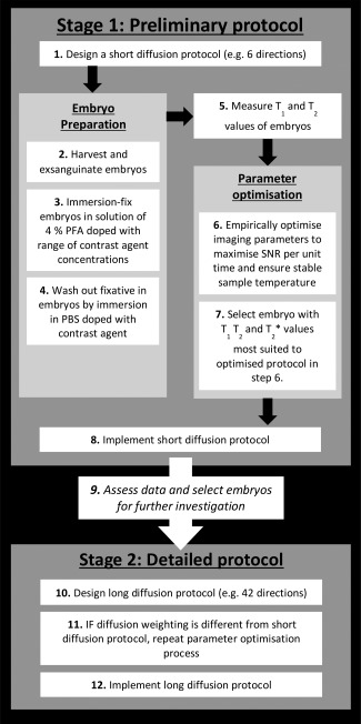
We developed two protocols for designing experimental procedures for diffusion μMRI of mouse embryos, which take into account the effect of embryo preparation and pulse sequence parameters on resulting data. We applied our protocols to an investigation of the splotch mouse model as an example implementation.
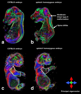
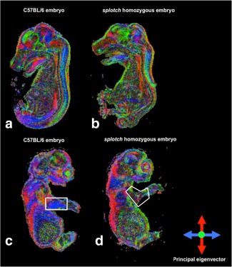
The protocols provide DTI data in 24 min per direction at 75 μm isotropic using a three-dimensional fast spin-echo sequence, enabling preliminary imaging in 3 h (6 directions plus one unweighted measurement), or detailed imaging in 9 h (42 directions plus six unweighted measurements). Application to the splotch model enabled assessment of spinal cord pathology.
We present guidelines for designing diffusion μMRI experiments, which may be adapted for different studies and research facilities. As they are suitable for routine use and may be readily implemented, we hope they will be adopted by the phenotyping community.
可视化新型组织对比度的先进方法对于对不断增加的突变小鼠胚胎进行表型分析至关重要。尽管扩散显微磁共振成像(μMRI)已用于胚胎表型分析,但由于扫描时间延长,其广泛的常规应用受到限制,并且尚无既定的实验程序可确保最佳数据采集。
我们开发了两种用于设计小鼠胚胎扩散μMRI实验程序的方案,该方案考虑了胚胎制备和脉冲序列参数对所得数据的影响。作为示例实施,我们将我们的方案应用于对斑点小鼠模型的研究。
该方案使用三维快速自旋回波序列在各向同性为75μm的情况下,每个方向在24分钟内提供扩散张量成像(DTI)数据,能够在3小时内进行初步成像(6个方向加一次无加权测量),或在9小时内进行详细成像(42个方向加六次无加权测量)。应用于斑点模型能够评估脊髓病理学。
我们提出了设计扩散μMRI实验的指南,该指南可适用于不同的研究和研究设施。由于它们适用于常规使用且易于实施,我们希望它们能被表型分析社区采用。