• 文献检索
  • 文档翻译
  • 深度研究
  • 学术资讯
  • Suppr Zotero 插件Zotero 插件
  • 邀请有礼
  • 套餐&价格
  • 历史记录
应用&插件
Suppr Zotero 插件Zotero 插件浏览器插件Mac 客户端Windows 客户端微信小程序
定价
高级版会员购买积分包购买API积分包
服务
文献检索文档翻译深度研究API 文档MCP 服务
关于我们
关于 Suppr公司介绍联系我们用户协议隐私条款
关注我们

Suppr 超能文献

核心技术专利:CN118964589B侵权必究
粤ICP备2023148730 号-1Suppr @ 2026

文献检索

告别复杂PubMed语法,用中文像聊天一样搜索,搜遍4000万医学文献。AI智能推荐,让科研检索更轻松。

立即免费搜索

文件翻译

保留排版,准确专业,支持PDF/Word/PPT等文件格式,支持 12+语言互译。

免费翻译文档

深度研究

AI帮你快速写综述,25分钟生成高质量综述,智能提取关键信息,辅助科研写作。

立即免费体验

非霍奇金淋巴瘤中的细胞增殖。Ki-67抗体染色的数字图像分析。

Cell proliferation in non-Hodgkin's lymphomas. Digital image analysis of Ki-67 antibody staining.

作者信息

Schwartz B R, Pinkus G, Bacus S, Toder M, Weinberg D S

机构信息

Department of Pathology, Brigham and Women's Hospital, Boston, Massachusetts 02115.

出版信息

Am J Pathol. 1989 Feb;134(2):327-36.

PMID:2464940
原文链接:https://pmc.ncbi.nlm.nih.gov/articles/PMC1879592/
Abstract

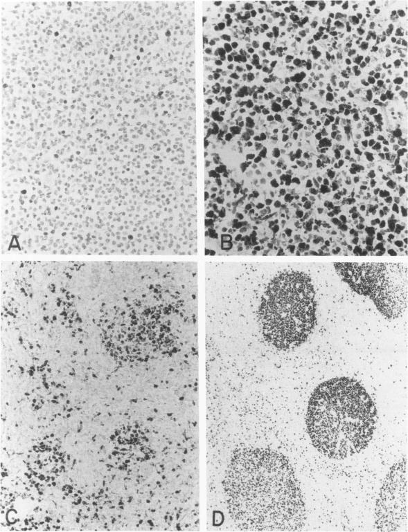
Ki-67 is a monoclonal antibody to a nuclear antigen present in cycling human cells but not in resting cells. The authors have performed immunoperoxidase on non-Hodgkin's lymphomas using Ki-67 antibody in order to correlate proliferation rates with tumor grade and type, and compare Ki-67 staining with S-phase content as determined by flow cytometry. Ki-67 staining of 109 sections was quantitated using a digital image analysis system (CAS 100). There was a significant difference among mean overall Ki-67 staining values in Working Formulation low (13.7%), intermediate (42.6%), and high grade tumors (57.9%, P less than 0.00001). The level of significance improved when a revised grading system was formulated based on proliferative activity, with the inclusion of diffuse large cell lymphomas in the high grade category. Within nodular and a few diffuse lymphomas, there were well-defined proliferation centers in which Ki-67 staining showed no correlation with grade. Flow cytometric DNA determination was performed on 74 specimens, and there was a positive correlation between Ki-67 positivity and S phase content (r = 0.66). It is concluded that Ki-67 staining of tissue sections is an alternative to flow cytometric quantitation of cell cycle activity in lymphomas, and provides the advantage of revealing histologic patterns of proliferation. By including G1 phase cells, Ki-67 staining allows a more complete determination of total cell cycle activity in lymphomas.

摘要

Ki-67是一种针对人类循环细胞中存在的核抗原的单克隆抗体,静止细胞中不存在该抗原。作者使用Ki-67抗体对非霍奇金淋巴瘤进行免疫过氧化物酶检测,以便将增殖率与肿瘤分级和类型相关联,并将Ki-67染色与通过流式细胞术测定的S期含量进行比较。使用数字图像分析系统(CAS 100)对109个切片的Ki-67染色进行定量。工作分类法中的低级别(13.7%)、中级别(42.6%)和高级别肿瘤(57.9%,P小于0.00001)的平均总体Ki-67染色值之间存在显著差异。当基于增殖活性制定修订的分级系统,将弥漫性大细胞淋巴瘤纳入高级别类别时,显著性水平有所提高。在结节性和少数弥漫性淋巴瘤中,存在明确的增殖中心,其中Ki-67染色与分级无关。对74个标本进行了流式细胞术DNA测定,Ki-67阳性与S期含量之间存在正相关(r = 0.66)。得出的结论是,组织切片的Ki-67染色是淋巴瘤细胞周期活性流式细胞术定量分析的一种替代方法,并且具有揭示增殖组织学模式的优势。通过纳入G1期细胞,Ki-67染色能够更全面地测定淋巴瘤中的总细胞周期活性。

https://cdn.ncbi.nlm.nih.gov/pmc/blobs/1343/1879592/2a565865730f/amjpathol00122-0101-a.jpg

相似文献

1
Cell proliferation in non-Hodgkin's lymphomas. Digital image analysis of Ki-67 antibody staining.非霍奇金淋巴瘤中的细胞增殖。Ki-67抗体染色的数字图像分析。
Am J Pathol. 1989 Feb;134(2):327-36.
2
Proliferation markers in non-Hodgkin's lymphoma. A comparative study between cytophotometric quantitation of Ki-67 and flow cytometric proliferation index on fine needle aspirates.非霍奇金淋巴瘤中的增殖标志物。细针穿刺抽吸物中Ki-67细胞光度法定量与流式细胞术增殖指数的比较研究。
Anal Quant Cytol Histol. 1993 Jun;15(3):179-86.
3
The role of cell cycle activity in the generation of morphologic heterogeneity in non-Hodgkin's lymphoma.细胞周期活性在非霍奇金淋巴瘤形态学异质性产生中的作用。
Am J Pathol. 1989 Oct;135(4):759-70.
4
MIB-1, Ki67, and PCNA scores and DNA flow cytometry in intermediate grade malignant lymphomas.中级别恶性淋巴瘤中的MIB-1、Ki67和增殖细胞核抗原评分及DNA流式细胞术
J Clin Pathol. 1994 Jan;47(1):18-22. doi: 10.1136/jcp.47.1.18.
5
Quantitative study of KI-67 antibody staining in non-Hodgkin's lymphomas using image analysis.
Anal Quant Cytol Histol. 1991 Aug;13(4):279-87.
6
Comparative assessment of proliferation and DNA content in breast carcinoma by image analysis and flow cytometry.通过图像分析和流式细胞术对乳腺癌增殖和DNA含量的比较评估
Am J Pathol. 1990 May;136(5):1115-24.
7
Biological heterogeneity of diffuse mixed small and large cell non-Hodgkin's lymphomas assessed by DNA flow cytometry and Ki67.通过DNA流式细胞术和Ki67评估弥漫性混合小细胞和大细胞非霍奇金淋巴瘤的生物学异质性。
Leuk Lymphoma. 1995 Nov;19(5-6):467-72. doi: 10.3109/10428199509112206.
8
The prognostic value of Ki-67 antigen in non-Hodgkin lymphoma of Waldeyer ring and the nasal cavity.
Cancer. 1992 Nov 1;70(9):2342-9. doi: 10.1002/1097-0142(19921101)70:9<2342::aid-cncr2820700922>3.0.co;2-6.
9
DNA image cytometry and the expression of proliferative markers (proliferating cell nuclear antigen and Ki67) in non-Hodgkin's lymphomas.非霍奇金淋巴瘤中的DNA图像细胞术及增殖标志物(增殖细胞核抗原和Ki67)的表达
Mod Pathol. 1995 Jan;8(1):51-8.
10
Analysis of proliferating cell fraction determined by monoclonal antibody to M1-subunit ribonucleotide reductase and Ki-67 in relation to p53 protein expression in fine-needle aspirates from non-Hodgkin's lymphomas.应用针对M1亚基核糖核苷酸还原酶的单克隆抗体和Ki-67对非霍奇金淋巴瘤细针穿刺抽吸物中增殖细胞分数进行分析,并与p53蛋白表达相关联。
Cytopathology. 2000 Oct;11(5):290-301. doi: 10.1046/j.1365-2303.2000.00234.x.

引用本文的文献

1
Comparison of Three Ki-67 Index Quantification Methods and Clinical Significance in Pancreatic Neuroendocrine Tumors.三种Ki-67指数定量方法在胰腺神经内分泌肿瘤中的比较及临床意义
Endocr Pathol. 2015 Sep;26(3):255-62. doi: 10.1007/s12022-015-9379-2.
2
P53, bcl-2, ki-67 li (labeling index) status in benign, proliferative, and malignant ovarian surface epithelial neoplasms.P53、bcl-2、ki-67 标记指数(LI)在良性、增殖性和恶性卵巢表面上皮性肿瘤中的状态。
Eurasian J Med. 2009 Apr;41(1):10-4.
3
An Efficient Computational Framework for the Analysis of Whole Slide Images: Application to Follicular Lymphoma Immunohistochemistry.

本文引用的文献

1
Proliferation kinetics of malignant non-Hodgkin's lymphomas related to histopathology of lymph node biopsies.与淋巴结活检组织病理学相关的恶性非霍奇金淋巴瘤的增殖动力学
Virchows Arch A Pathol Anat Histol. 1980;389(3):397-407. doi: 10.1007/BF00430662.
2
The pre-treatment proliferative activity of non-Hodgkin's lymphoma cells.
Eur J Cancer (1965). 1981 Jan;17(1):99-108. doi: 10.1016/0014-2964(81)90218-8.
3
Flow cytometry in the diagnosis and classification of malignant lymphoma and leukemia.流式细胞术在恶性淋巴瘤和白血病的诊断与分类中的应用
一种用于全切片图像分析的高效计算框架:在滤泡性淋巴瘤免疫组织化学中的应用。
J Comput Sci. 2012 Sep 1;3(5):269-279. doi: 10.1016/j.jocs.2012.01.009. Epub 2012 Mar 6.
4
Combined proliferating cell nuclear antigen and morphometric analysis in the diagnosis of cutaneous lymphoid infiltrates.增殖细胞核抗原与形态计量分析相结合在皮肤淋巴细胞浸润诊断中的应用
J Clin Pathol. 1993 Feb;46(2):129-34. doi: 10.1136/jcp.46.2.129.
5
Computer-aided image analysis applied to immunogold-silver staining: evaluation of proliferating cell nuclear antigen (PCNA)-reactive sites in paraffin sections.应用于免疫金银染色的计算机辅助图像分析:石蜡切片中增殖细胞核抗原(PCNA)反应位点的评估
Histochemistry. 1994 Sep;102(3):177-81. doi: 10.1007/BF00268894.
6
Biological grading of breast cancer using antibodies to proliferating cells and other markers.使用针对增殖细胞及其他标志物的抗体对乳腺癌进行生物学分级。
Am J Pathol. 1989 Nov;135(5):783-92.
7
Proliferation in non-Hodgkin's lymphoma: a comparison of Ki-67 staining on fine needle aspiration and cryostat sections.非霍奇金淋巴瘤中的增殖:细针穿刺抽吸物与冷冻切片上Ki-67染色的比较
J Clin Pathol. 1990 Apr;43(4):325-8. doi: 10.1136/jcp.43.4.325.
8
Review: assessment of cell proliferation in histological material.综述:组织学材料中细胞增殖的评估
J Clin Pathol. 1990 Mar;43(3):184-92. doi: 10.1136/jcp.43.3.184.
9
How malignant is malignant? A brief review of the microscopic assessment of human neoplasms, and the prediction of whether they will metastasize and kill.
Clin Exp Metastasis. 1991 Mar-Apr;9(2):127-37. doi: 10.1007/BF01756384.
10
Proliferation markers Ki-67 and p105 in soft-tissue lesions. Correlation with DNA flow cytometric characteristics.软组织病变中的增殖标志物Ki-67和p105。与DNA流式细胞术特征的相关性。
Am J Pathol. 1990 Dec;137(6):1491-500.
Cancer. 1982 Sep 15;50(6):1122-35. doi: 10.1002/1097-0142(19820915)50:6<1122::aid-cncr2820500616>3.0.co;2-q.
4
Morphologic criteria for the differentiation of follicular lymphoma from florid reactive follicular hyperplasia: a study of 80 cases.
Cancer. 1981 Oct 15;48(8):1794-806. doi: 10.1002/1097-0142(19811015)48:8<1794::aid-cncr2820480817>3.0.co;2-m.
5
Percentage of cells in the S phase of the cell cycle in human lymphoma determined by flow cytometry.通过流式细胞术测定的人类淋巴瘤细胞周期S期细胞的百分比。
Cytometry. 1980 Nov;1(3):171-4. doi: 10.1002/cyto.990010302.
6
Spontaneous (H)-thymidine uptake in histological subgroups of human B-cell lymphomas.
Scand J Haematol. 1981 Mar;26(3):221-34. doi: 10.1111/j.1600-0609.1981.tb01650.x.
7
Labeling index as a prognostic marker in non-Hodgkin's lymphomas.标记指数作为非霍奇金淋巴瘤的预后标志物
J Natl Cancer Inst. 1981 Jan;66(1):1-5.
8
Lymphoblastic lymphoma: a clinicopathologic study of 95 patients.淋巴母细胞淋巴瘤:95例患者的临床病理研究
Cancer. 1981 Dec 1;48(11):2347-57. doi: 10.1002/1097-0142(19811201)48:11<2347::aid-cncr2820481102>3.0.co;2-x.
9
Malignant lymphoma, intermediate lymphocytic type: a clinicopathologic study of 42 cases.恶性淋巴瘤,中间淋巴细胞型:42例临床病理研究
Cancer. 1981 Sep 15;48(6):1415-25. doi: 10.1002/1097-0142(19810915)48:6<1415::aid-cncr2820480625>3.0.co;2-n.
10
The biology of tumor growth in the non-Hodgkin's lymphomas. A dual parameter flow cytometry study of 220 cases.非霍奇金淋巴瘤中肿瘤生长的生物学特性。220例病例的双参数流式细胞术研究。
J Clin Invest. 1984 Apr;73(4):1201-14. doi: 10.1172/JCI111306.