Bacus S S, Goldschmidt R, Chin D, Moran G, Weinberg D, Bacus J W
Cell Analysis Systems, Inc., Lombard, IL 60148.
Am J Pathol. 1989 Nov;135(5):783-92.
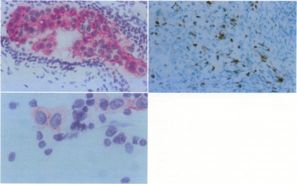
Quantitation of immunohistochemical staining by image analysis was performed on 50 breast cancers stained with the monoclonal antibody Ki-67 to determine the growth fraction and its correlation with tumor grade. A high degree of correlation was shown. For each case the DNA ploidy was determined by quantitation of the DNA Feulgen stain by computerized microdensitometry. DNA content of breast tumor cells correlated to the histopathologic grade at which poorly differentiated tumors are more likely to be aneuploid. Quantitation of immunohistochemistry for estrogen and progesterone receptors had a high degree of correlation with the steroid binding assay, such as dextran-coated charcoal assay (DCCA), and were weakly correlated to histologic grade. In summary, our results indicated that quantitation of Ki-67-positive nuclear area and of DNA content by image analysis provides an objective method for assessing tumor cell growth fraction and DNA ploidy. Quantitation of steroid receptors by immunohistochemistry is a better and easier technique than those currently used to determine the best therapy for postmenopausal women. These methods can be performed on small frozen sections or needle aspirates in quantities that are insufficient for current steroid binding assays. Thus, this method is prognosticly useful even for patients with small breast lesions.
通过图像分析对50例用单克隆抗体Ki-67染色的乳腺癌进行免疫组化染色定量,以确定生长分数及其与肿瘤分级的相关性。结果显示存在高度相关性。对每例病例,通过计算机化显微密度测定法对DNA Feulgen染色进行定量来确定DNA倍体。乳腺肿瘤细胞的DNA含量与组织病理学分级相关,其中低分化肿瘤更可能为非整倍体。雌激素和孕激素受体免疫组化定量与类固醇结合测定法(如葡聚糖包被活性炭测定法,DCCA)高度相关,与组织学分级弱相关。总之,我们的结果表明,通过图像分析对Ki-67阳性核面积和DNA含量进行定量,为评估肿瘤细胞生长分数和DNA倍体提供了一种客观方法。免疫组化定量类固醇受体是一种比目前用于确定绝经后女性最佳治疗方法的技术更好、更简便的技术。这些方法可在小的冰冻切片或针吸物上进行,而这些样本量对于目前的类固醇结合测定来说是不足的。因此,即使对于小乳腺病变患者,该方法在预后方面也很有用。