• 文献检索
  • 文档翻译
  • 深度研究
  • 学术资讯
  • Suppr Zotero 插件Zotero 插件
  • 邀请有礼
  • 套餐&价格
  • 历史记录
应用&插件
Suppr Zotero 插件Zotero 插件浏览器插件Mac 客户端Windows 客户端微信小程序
定价
高级版会员购买积分包购买API积分包
服务
文献检索文档翻译深度研究API 文档MCP 服务
关于我们
关于 Suppr公司介绍联系我们用户协议隐私条款
关注我们

Suppr 超能文献

核心技术专利:CN118964589B侵权必究
粤ICP备2023148730 号-1Suppr @ 2026

文献检索

告别复杂PubMed语法,用中文像聊天一样搜索,搜遍4000万医学文献。AI智能推荐,让科研检索更轻松。

立即免费搜索

文件翻译

保留排版,准确专业,支持PDF/Word/PPT等文件格式,支持 12+语言互译。

免费翻译文档

深度研究

AI帮你快速写综述,25分钟生成高质量综述,智能提取关键信息,辅助科研写作。

立即免费体验

基于心脏电影磁共振成像的左心室自动定位:一种新的基于频谱的计算机辅助工具。

Automatic localization of the left ventricle from cardiac cine magnetic resonance imaging: a new spectrum-based computer-aided tool.

作者信息

Zhong Liang, Zhang Jun-Mei, Zhao Xiaodan, Tan Ru San, Wan Min

机构信息

Bioengineering Department, National Heart Centre Singapore, Singapore, Singapore; Cardiovascular & Metabolic Disorders Program, Duke-NUS Graduate Medical School Singapore, Singapore, Singapore.

Bioengineering Department, National Heart Centre Singapore, Singapore, Singapore.

出版信息

PLoS One. 2014 Apr 10;9(4):e92382. doi: 10.1371/journal.pone.0092382. eCollection 2014.

DOI:10.1371/journal.pone.0092382
PMID:24722328
原文链接:https://pmc.ncbi.nlm.nih.gov/articles/PMC3982962/
Abstract

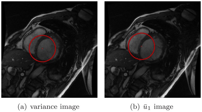
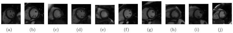
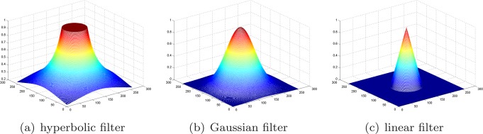
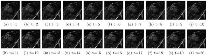
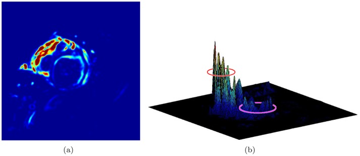
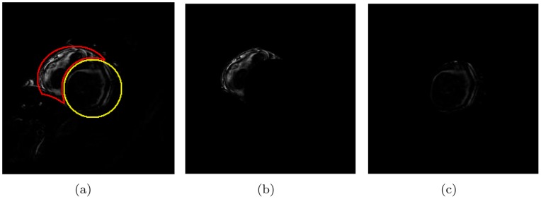
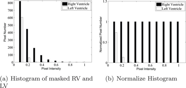
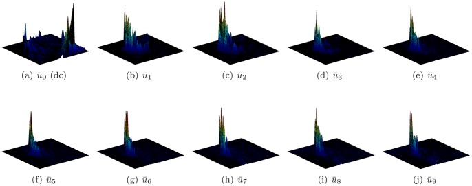
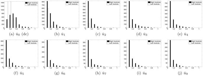
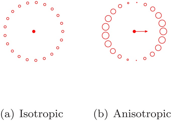
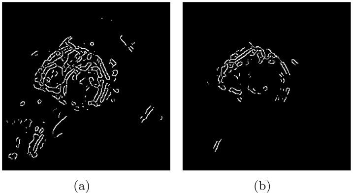
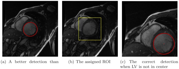
Traditionally, cardiac image analysis is done manually. Automatic image processing can help with the repetitive tasks, and also deal with huge amounts of data, a task which would be humanly tedious. This study aims to develop a spectrum-based computer-aided tool to locate the left ventricle using images obtained via cardiac magnetic resonance imaging. Discrete Fourier Transform was conducted pixelwise on the image sequence. Harmonic images of all frequencies were analyzed visually and quantitatively to determine different patterns of the left and right ventricles on spectrum. The first and fifth harmonic images were selected to perform an anisotropic weighted circle Hough detection. This tool was then tested in ten volunteers. Our tool was able to locate the left ventricle in all cases and had a significantly higher cropping ratio of 0.165 than did earlier studies. In conclusion, a new spectrum-based computer aided tool has been proposed and developed for automatic left ventricle localization. The development of this technique, which will enable the automatic location and further segmentation of the left ventricle, will have a significant impact in research and in diagnostic settings. We envisage that this automated method could be used by radiographers and cardiologists to diagnose and assess ventricular function in patients with diverse heart diseases.

摘要

传统上,心脏图像分析是通过人工完成的。自动图像处理有助于完成重复性任务,还能处理大量数据,而这是一项人工操作会很繁琐的任务。本研究旨在开发一种基于频谱的计算机辅助工具,利用通过心脏磁共振成像获得的图像来定位左心室。对图像序列逐像素进行离散傅里叶变换。对所有频率的谐波图像进行视觉和定量分析,以确定频谱上左心室和右心室的不同模式。选择第一和第五谐波图像进行各向异性加权圆霍夫检测。然后在十名志愿者身上对该工具进行测试。我们的工具在所有情况下都能够定位左心室,并且裁剪率为0.165,明显高于早期研究。总之,已经提出并开发了一种新的基于频谱的计算机辅助工具用于自动左心室定位。这项技术的发展将能够实现左心室的自动定位和进一步分割,对研究和诊断环境将产生重大影响。我们设想,这种自动化方法可由放射技师和心脏病专家用于诊断和评估患有各种心脏病患者的心室功能。

https://cdn.ncbi.nlm.nih.gov/pmc/blobs/f9f3/3982962/6f9bc494b4b8/pone.0092382.g015.jpg
https://cdn.ncbi.nlm.nih.gov/pmc/blobs/f9f3/3982962/bb4636c25636/pone.0092382.g001.jpg
https://cdn.ncbi.nlm.nih.gov/pmc/blobs/f9f3/3982962/9a8931e3dd33/pone.0092382.g002.jpg
https://cdn.ncbi.nlm.nih.gov/pmc/blobs/f9f3/3982962/9fc49311d5c9/pone.0092382.g003.jpg
https://cdn.ncbi.nlm.nih.gov/pmc/blobs/f9f3/3982962/68e8f64768b9/pone.0092382.g004.jpg
https://cdn.ncbi.nlm.nih.gov/pmc/blobs/f9f3/3982962/937142a7313a/pone.0092382.g005.jpg
https://cdn.ncbi.nlm.nih.gov/pmc/blobs/f9f3/3982962/4ec48e1a23a3/pone.0092382.g006.jpg
https://cdn.ncbi.nlm.nih.gov/pmc/blobs/f9f3/3982962/678d8aaaa845/pone.0092382.g007.jpg
https://cdn.ncbi.nlm.nih.gov/pmc/blobs/f9f3/3982962/926aa8435646/pone.0092382.g008.jpg
https://cdn.ncbi.nlm.nih.gov/pmc/blobs/f9f3/3982962/1fc311640652/pone.0092382.g009.jpg
https://cdn.ncbi.nlm.nih.gov/pmc/blobs/f9f3/3982962/89e50bcbe6e0/pone.0092382.g010.jpg
https://cdn.ncbi.nlm.nih.gov/pmc/blobs/f9f3/3982962/13d65781bad0/pone.0092382.g011.jpg
https://cdn.ncbi.nlm.nih.gov/pmc/blobs/f9f3/3982962/e66f84f1eb9b/pone.0092382.g012.jpg
https://cdn.ncbi.nlm.nih.gov/pmc/blobs/f9f3/3982962/05ed70a54434/pone.0092382.g013.jpg
https://cdn.ncbi.nlm.nih.gov/pmc/blobs/f9f3/3982962/3ada5fccdb9a/pone.0092382.g014.jpg
https://cdn.ncbi.nlm.nih.gov/pmc/blobs/f9f3/3982962/6f9bc494b4b8/pone.0092382.g015.jpg

相似文献

1
Automatic localization of the left ventricle from cardiac cine magnetic resonance imaging: a new spectrum-based computer-aided tool.基于心脏电影磁共振成像的左心室自动定位:一种新的基于频谱的计算机辅助工具。
PLoS One. 2014 Apr 10;9(4):e92382. doi: 10.1371/journal.pone.0092382. eCollection 2014.
2
Automatic left ventricle segmentation in cardiac MRI using topological stable-state thresholding and region restricted dynamic programming.基于拓扑稳定状态阈值法和区域受限动态规划的心脏 MRI 自动左心室分割。
Acad Radiol. 2012 Jun;19(6):723-31. doi: 10.1016/j.acra.2012.02.011. Epub 2012 Apr 1.
3
Hybrid segmentation of left ventricle in cardiac MRI using Gaussian-mixture model and region restricted dynamic programming.基于高斯混合模型和区域受限动态规划的心脏 MRI 左心室混合分割。
Magn Reson Imaging. 2013 May;31(4):575-84. doi: 10.1016/j.mri.2012.10.004. Epub 2012 Dec 14.
4
An image-based comprehensive approach for automatic segmentation of left ventricle from cardiac short axis cine MR images.基于图像的综合方法用于自动分割心脏短轴电影磁共振图像中的左心室。
J Digit Imaging. 2011 Aug;24(4):598-608. doi: 10.1007/s10278-010-9315-4.
5
Automatic recovery of the left ventricular blood pool in cardiac cine MR images.心脏电影磁共振图像中左心室血池的自动恢复
Med Image Comput Comput Assist Interv. 2008;11(Pt 1):110-8. doi: 10.1007/978-3-540-85988-8_14.
6
Fully automated segmentation of the left ventricle in cine cardiac MRI using neural network regression.使用神经网络回归对心脏电影磁共振成像中的左心室进行全自动分割。
J Magn Reson Imaging. 2018 Jul;48(1):140-152. doi: 10.1002/jmri.25932. Epub 2018 Jan 9.
7
Evaluation of cardiac biventricular segmentation from multiaxis MRI data: a multicenter study.基于多轴磁共振成像数据的心脏双心室分割评估:一项多中心研究
J Magn Reson Imaging. 2008 Sep;28(3):626-36. doi: 10.1002/jmri.21520.
8
Fully Automatic initialization and segmentation of left and right ventricles for large-scale cardiac MRI using a deeply supervised network and 3D-ASM.基于深度监督网络和 3D-ASM 的大规模心脏 MRI 左、右心室全自动初始化和分割。
Comput Methods Programs Biomed. 2023 Oct;240:107679. doi: 10.1016/j.cmpb.2023.107679. Epub 2023 Jun 14.
9
Automatic image-driven segmentation of the ventricles in cardiac cine MRI.心脏电影磁共振成像中心室的自动图像驱动分割
J Magn Reson Imaging. 2008 Aug;28(2):366-74. doi: 10.1002/jmri.21451.
10
Automatic cardiac LV segmentation in MRI using modified graph cuts with smoothness and interslice constraints.基于具有平滑度和层间约束的改进图割算法在心脏磁共振成像中自动分割左心室。
Magn Reson Med. 2014 Dec;72(6):1775-84. doi: 10.1002/mrm.25079. Epub 2013 Dec 17.

引用本文的文献

1
Left Ventricle Detection from Cardiac Magnetic Resonance Relaxometry Images Using Visual Transformer.基于视觉Transformer的心脏磁共振弛豫定量图像左心室检测。
Sensors (Basel). 2023 Mar 21;23(6):3321. doi: 10.3390/s23063321.
2
Fully Automated 3D Cardiac MRI Localisation and Segmentation Using Deep Neural Networks.使用深度神经网络的全自动3D心脏磁共振成像定位与分割
J Imaging. 2020 Jul 6;6(7):65. doi: 10.3390/jimaging6070065.
3
Artificial intelligence in pediatric and adult congenital cardiac MRI: an unmet clinical need.人工智能在儿科和成人先天性心脏病磁共振成像中的应用:一项未满足的临床需求。

本文引用的文献

1
Myocardial contractile dysfunction associated with increased 3-month and 1-year mortality in hospitalized patients with heart failure and preserved ejection fraction.心力衰竭射血分数保留患者住院期间心肌收缩功能障碍与 3 个月和 1 年死亡率增加相关。
Int J Cardiol. 2013 Oct 3;168(3):1975-83. doi: 10.1016/j.ijcard.2012.12.084. Epub 2013 Jan 19.
2
Right ventricular regional wall curvedness and area strain in patients with repaired tetralogy of Fallot.右心室局部壁弯曲度和面积应变在法洛四联症修复患者中的变化。
Am J Physiol Heart Circ Physiol. 2012 Mar 15;302(6):H1306-16. doi: 10.1152/ajpheart.00679.2011. Epub 2011 Dec 30.
3
Cardiovasc Diagn Ther. 2019 Oct;9(Suppl 2):S310-S325. doi: 10.21037/cdt.2019.06.09.
4
A Deep Learning Segmentation Approach in Free-Breathing Real-Time Cardiac Magnetic Resonance Imaging.基于自由呼吸实时心脏磁共振成像的深度学习分割方法。
Biomed Res Int. 2019 Jul 30;2019:5636423. doi: 10.1155/2019/5636423. eCollection 2019.
5
Automatic localization of the left ventricular blood pool centroid in short axis cardiac cine MR images.短轴心脏电影磁共振图像中左心室血池质心的自动定位。
Med Biol Eng Comput. 2018 Jun;56(6):1053-1062. doi: 10.1007/s11517-017-1750-7. Epub 2017 Nov 17.
6
Convolutional Neural Network for the Detection of End-Diastole and End-Systole Frames in Free-Breathing Cardiac Magnetic Resonance Imaging.用于自由呼吸心脏磁共振成像中舒张末期和收缩末期帧检测的卷积神经网络
Comput Math Methods Med. 2017;2017:1640835. doi: 10.1155/2017/1640835. Epub 2017 Jul 26.
7
The electronic stethoscope.电子听诊器。
Biomed Eng Online. 2015 Jul 10;14:66. doi: 10.1186/s12938-015-0056-y.
8
The electronic health record for translational research.用于转化研究的电子健康记录。
J Cardiovasc Transl Res. 2014 Aug;7(6):607-14. doi: 10.1007/s12265-014-9579-z. Epub 2014 Jul 29.
Attenuation of stress-based ventricular contractility in patients with heart failure and normal ejection fraction.
心力衰竭和射血分数正常患者的基于压力的心室收缩力衰减。
Ann Acad Med Singap. 2011 Apr;40(4):179-85.
4
Impact of surgical ventricular restoration on ventricular shape, wall stress, and function in heart failure patients.外科心室修复对心力衰竭患者心室形态、壁应力和功能的影响。
Am J Physiol Heart Circ Physiol. 2011 May;300(5):H1653-60. doi: 10.1152/ajpheart.00021.2011. Epub 2011 Feb 25.
5
A review of segmentation methods in short axis cardiac MR images.短轴心脏磁共振图像分割方法综述。
Med Image Anal. 2011 Apr;15(2):169-84. doi: 10.1016/j.media.2010.12.004. Epub 2010 Dec 24.
6
Total variation with automatic hyper-parameter estimation.具有自动超参数估计的总变差
Annu Int Conf IEEE Eng Med Biol Soc. 2008;2008:443-6. doi: 10.1109/IEMBS.2008.4649185.
7
Left ventricular regional wall curvedness and wall stress in patients with ischemic dilated cardiomyopathy.缺血性扩张型心肌病患者的左心室局部壁曲率和壁应力
Am J Physiol Heart Circ Physiol. 2009 Mar;296(3):H573-84. doi: 10.1152/ajpheart.00525.2008. Epub 2009 Jan 2.
8
Automatic recovery of the left ventricular blood pool in cardiac cine MR images.心脏电影磁共振图像中左心室血池的自动恢复
Med Image Comput Comput Assist Interv. 2008;11(Pt 1):110-8. doi: 10.1007/978-3-540-85988-8_14.
9
Snakes, shapes, and gradient vector flow.蛇形、形状与梯度向量流。
IEEE Trans Image Process. 1998;7(3):359-69. doi: 10.1109/83.661186.
10
Robust shape tracking with multiple models in ultrasound images.超声图像中基于多模型的稳健形状跟踪
IEEE Trans Image Process. 2008 Mar;17(3):392-406. doi: 10.1109/TIP.2007.915552.