Department of Biochemistry and Molecular Biophysics, and Department of Physiology and Cellular Biophysics, Columbia University, New York, NY 10032, USA.
Q Rev Biophys. 2014 Feb;47(1):49-93. doi: 10.1017/S0033583514000018.
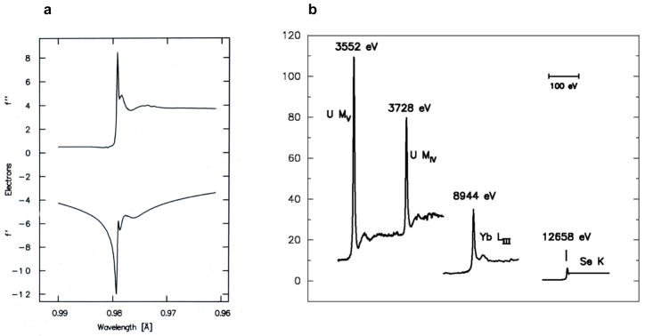
X-ray diffraction patterns from crystals of biological macromolecules contain sufficient information to define atomic structures, but atomic positions are inextricable without having electron-density images. Diffraction measurements provide amplitudes, but the computation of electron density also requires phases for the diffracted waves. The resonance phenomenon known as anomalous scattering offers a powerful solution to this phase problem. Exploiting scattering resonances from diverse elements, the methods of MAD (multiwavelength anomalous diffraction) and SAD (single-wavelength anomalous diffraction) now predominate for de novo determinations of atomic-level biological structures. This review describes the physical underpinnings of anomalous diffraction methods, the evolution of these methods to their current maturity, the elements, procedures and instrumentation used for effective implementation, and the realm of applications.
生物大分子晶体的 X 射线衍射图谱包含足以定义原子结构的信息,但如果没有电子密度图像,原子位置就无法确定。衍射测量提供了振幅,但计算电子密度还需要衍射波的相位。被称为反常散射的共振现象为解决这个相位问题提供了有力的解决方案。利用来自不同元素的散射共振,MAD(多波长反常衍射)和 SAD(单波长反常衍射)方法现在在从头确定原子水平生物结构方面占据主导地位。本文综述了反常衍射方法的物理基础、这些方法的发展及其目前的成熟程度、有效实施所使用的元素、程序和仪器以及应用领域。