Kroidl Arne, Kroidl Inge, Bretzel Gisela, Löscher Thomas
Division of Infectious Diseases and Tropical Medicine, Medical Center of the University of Munich, Leopoldstr, 5, 80802 Munich, Germany.
BMC Infect Dis. 2014 Apr 16;14:206. doi: 10.1186/1471-2334-14-206.
The prevalence of Old World Cutaneous Leishmaniasis in the Mediterranean region is increasing and in Southern Europe often caused by Leishmania infantum. Spontaneous healing of cutaneous leishmaniasis is commonly observed, especially if caused by L. major, whereas L. infantum associated lesions have been reported with longer disease duration and decreased tendency for self-limitation, however, available information is sparse.
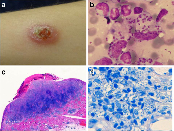
We report the case of an otherwise healthy woman from Southern Spain who presented with a seven years persistent, non-healing, painless, central ulcerated, nodular cutaneous lesion with a diameter of 2 cm of the forearm. Cutaneous leishmaniasis was diagnosed by smear and histology, showing large amounts of leishmania amastigotes in subepidermal histiocytes and extensive lymphocyte and plasma cell inflammation. L. infantum as the causative pathogen was confirmed by restriction fragment length polymorphism and microsatellite-PCR. Systemic or visceral involvement was excluded by negative leishmania serology and clinical presentation, relevant concomitant diseases or immunosuppression were excluded including quantification of immunoglobulin levels and lymphocyte phenotyping. Topical and systemic anti-infectious treatment options, often limited in terms of efficacy, tolerability and long lasting treatment duration, were considered. Treatment was successfully performed by surgical extraction in local anaesthesia only.
To our knowledge this is the longest reported duration of a L. infantum associated cutaneous leishmaniasis indicating a potential long lasting natural evolution of the disease in an otherwise healthy and immunocompetent patient, however, high parasite density may have reflected a lack of a L. infantum specific immune response. Complete surgical extraction can be successfully performed as treatment.
地中海地区旧世界皮肤利什曼病的患病率正在上升,在南欧通常由婴儿利什曼原虫引起。皮肤利什曼病通常可自愈,尤其是由大型利什曼原虫引起时,而据报道,与婴儿利什曼原虫相关的病变病程较长且自我限制倾向降低,然而,现有信息稀少。
我们报告了一例来自西班牙南部的健康女性病例,她的前臂出现了一个持续7年的、不愈合、无痛、中央溃疡、直径2厘米的结节性皮肤病变。通过涂片和组织学诊断为皮肤利什曼病,显示表皮下组织细胞中有大量利什曼原虫无鞭毛体,以及广泛的淋巴细胞和浆细胞炎症。通过限制性片段长度多态性和微卫星-PCR确认致病病原体为婴儿利什曼原虫。通过阴性利什曼原虫血清学和临床表现排除了全身或内脏受累,排除了相关的伴随疾病或免疫抑制,包括免疫球蛋白水平定量和淋巴细胞表型分析。考虑了局部和全身抗感染治疗方案,这些方案在疗效、耐受性和长期治疗持续时间方面往往有限。仅通过局部麻醉下的手术切除成功进行了治疗。
据我们所知,这是报道的与婴儿利什曼原虫相关的皮肤利什曼病最长病程,表明在一名原本健康且免疫功能正常的患者中,该疾病可能有潜在的长期自然演变,然而,高寄生虫密度可能反映了缺乏针对婴儿利什曼原虫的特异性免疫反应。完整的手术切除可作为成功的治疗方法。