• 文献检索
  • 文档翻译
  • 深度研究
  • 学术资讯
  • Suppr Zotero 插件Zotero 插件
  • 邀请有礼
  • 套餐&价格
  • 历史记录
应用&插件
Suppr Zotero 插件Zotero 插件浏览器插件Mac 客户端Windows 客户端微信小程序
定价
高级版会员购买积分包购买API积分包
服务
文献检索文档翻译深度研究API 文档MCP 服务
关于我们
关于 Suppr公司介绍联系我们用户协议隐私条款
关注我们

Suppr 超能文献

核心技术专利:CN118964589B侵权必究
粤ICP备2023148730 号-1Suppr @ 2026

文献检索

告别复杂PubMed语法,用中文像聊天一样搜索,搜遍4000万医学文献。AI智能推荐,让科研检索更轻松。

立即免费搜索

文件翻译

保留排版,准确专业,支持PDF/Word/PPT等文件格式,支持 12+语言互译。

免费翻译文档

深度研究

AI帮你快速写综述,25分钟生成高质量综述,智能提取关键信息,辅助科研写作。

立即免费体验

可靠且耐用的人类尸检和实验动物脑组织高尔基染色。

Reliable and durable Golgi staining of brain tissue from human autopsies and experimental animals.

机构信息

New York State Psychiatric Institute and Columbia University, Unit 42, 1051 Riverside Drive, New York, NY 10032, USA; Macedonian Academy of Sciences and Arts (MASA), Bul. Krste Petkov Misirkov 2, Skopje 1000, Macedonia; School of Medicine, University Ss. Cyril & Methodius, Vodnjanska 17, Skopje 1000, Macedonia.

Institute of Chemistry, Faculty of Natural Sciences and Mathematics, University Ss. Cyril & Methodius, Arhimedova 5, Skopje 1000, Macedonia.

出版信息

J Neurosci Methods. 2014 Jun 15;230:20-9. doi: 10.1016/j.jneumeth.2014.04.006. Epub 2014 Apr 18.

DOI:10.1016/j.jneumeth.2014.04.006
PMID:24747874
原文链接:https://pmc.ncbi.nlm.nih.gov/articles/PMC4080906/
Abstract

BACKGROUND

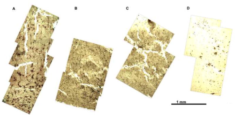
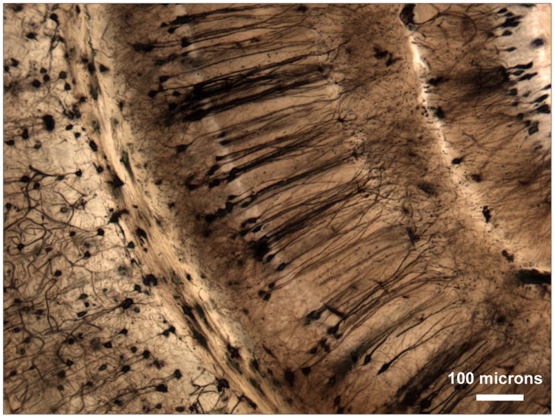
Golgi stains are notoriously capricious, particularly when applied to human brain. The well-known difficulties, which include complete failure of impregnation, patchy staining, unstable staining, and extensive crystalline deposits in superficial sections, have discouraged many from attempting to use these techniques. A reliable method that produces uniform impregnation in tissue from human autopsies and experimental animals is needed.

NEW METHOD

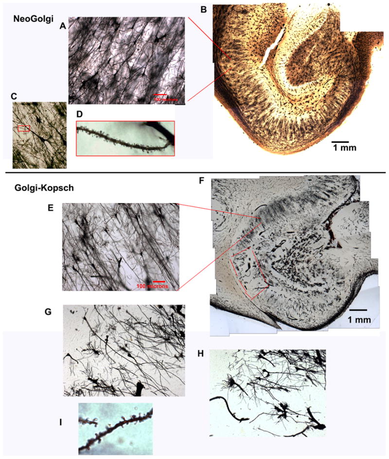
The method described, "NeoGolgi", modifies previous Golgi-Cox protocols (Glaser and Van der Loos, 1981). Changes include: much longer time (>10 weeks) in Golgi solution, agitation on a slowly rocking platform, more gradual infiltration with Parlodion, more thorough removal of excess staining solution during embedding, and shorter exposure to ammonia after infiltration.

RESULTS

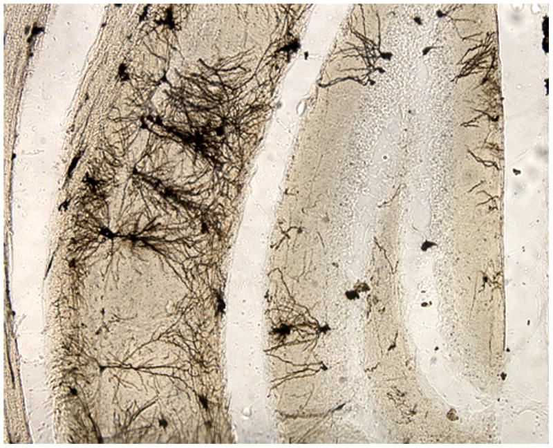
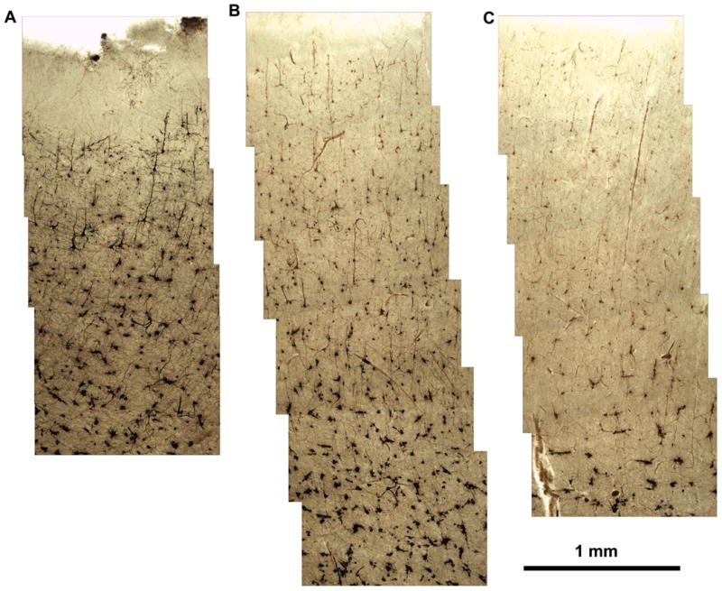
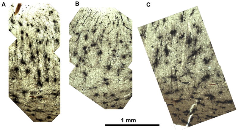
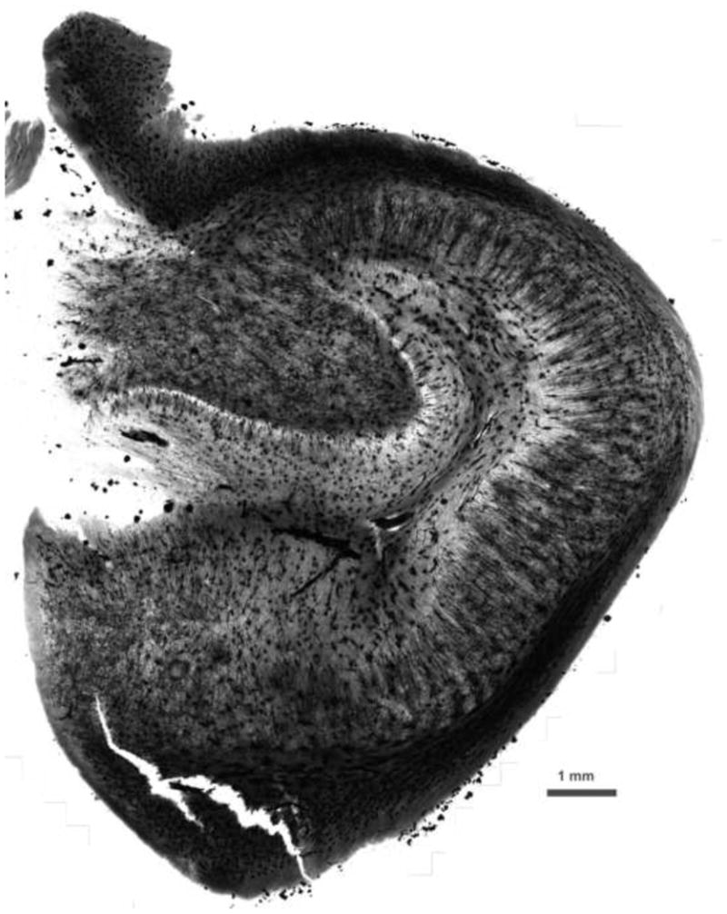
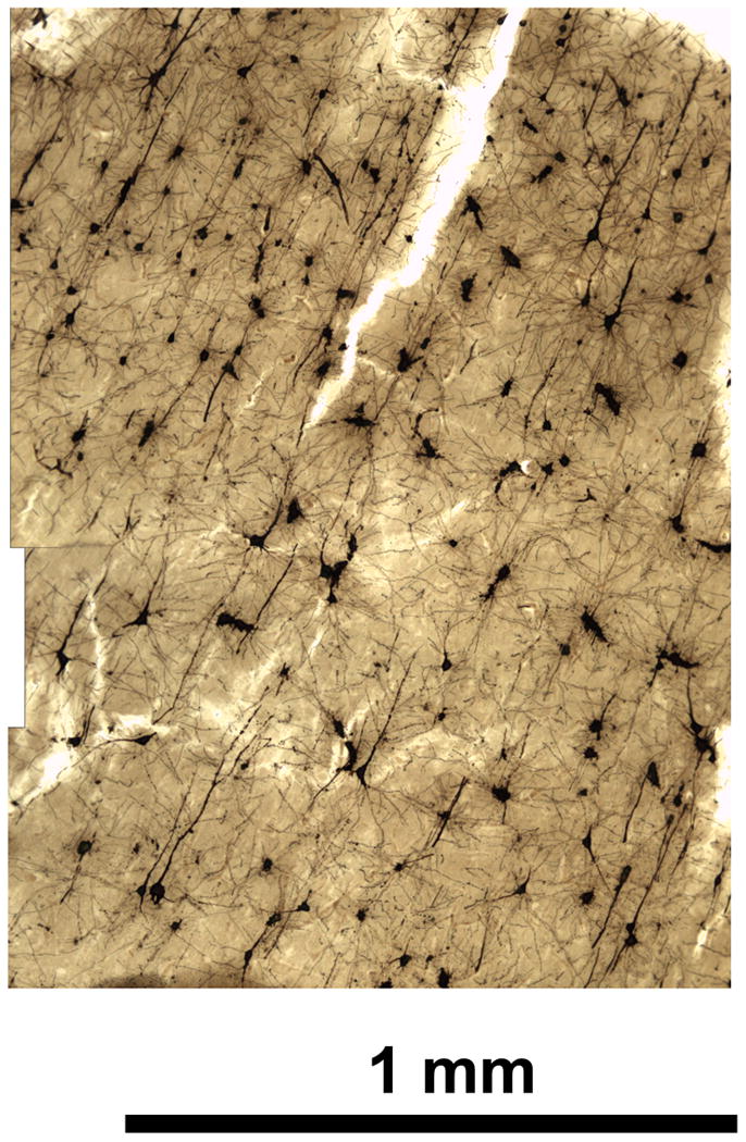
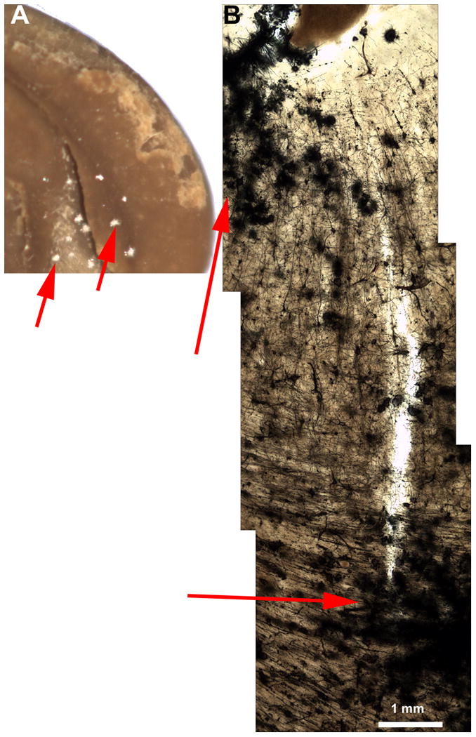
The procedure has successfully stained over 220 consecutive frontal or hippocampal blocks from more than 175 consecutive human autopsy cases. Dendritic spines are easily recognized, and background is clear, allowing examination of very thick (200 μm) sections. Stained neurons are evenly distributed within cortical regions. The stain is stable for at least eight years. Most importantly, all stained neurons are apparently well-impregnated, eliminating ambiguity between pathology and poor impregnation that is inherent to other methods.

COMPARISON WITH EXISTING METHODS

Most methods of Golgi staining are poorly predictable. They often fail completely, staining is patchy, and abnormal morphology is often indistinguishable from poor impregnation. "NeoGolgi" overcomes these problems.

CONCLUSION

Starting with unfixed tissue, it is possible to obtain Golgi staining of predictably high quality in brains from human autopsies and experimental animals.

摘要

背景

高尔基染色法非常不稳定,尤其是在应用于人脑时。众所周知,这种技术存在许多困难,包括完全浸渍失败、染色不均匀、染色不稳定以及在浅层切片中出现大量结晶沉积物等问题,这使得许多人望而却步,不愿尝试使用这些技术。目前需要一种可靠的方法,能够在人体和实验动物的组织中产生均匀的浸渍效果。

新方法

本文描述的方法“NeoGolgi”对以前的高尔基-考克斯(Golgi-Cox)方案进行了修改(Glaser 和 Van der Loos,1981)。更改包括:在高尔基溶液中浸泡时间更长(>10 周)、在缓慢晃动的平台上搅拌、用 Parlodion 更渐进地渗透、在包埋过程中更彻底地去除多余的染色溶液以及在渗透后更短时间暴露于氨水中。

结果

该程序已成功对 175 例连续尸检案例中的 220 个连续额或海马体块进行了染色。树突棘很容易识别,背景清晰,允许检查非常厚(200μm)的切片。染色神经元在皮质区域内均匀分布。该染色稳定至少八年。最重要的是,所有染色的神经元显然都被很好地浸渍,消除了其他方法中固有的病理学和浸渍不良之间的歧义。

与现有方法的比较

大多数高尔基染色方法都难以预测。它们经常完全失败,染色不均匀,异常形态学通常与浸渍不良难以区分。“NeoGolgi”克服了这些问题。

结论

从未固定的组织开始,有可能在人体和实验动物的大脑中获得可预测的高质量高尔基染色。

https://cdn.ncbi.nlm.nih.gov/pmc/blobs/fa41/4080906/4a6a9f5a67bc/nihms592867f9.jpg
https://cdn.ncbi.nlm.nih.gov/pmc/blobs/fa41/4080906/13a29fd61856/nihms592867f1.jpg
https://cdn.ncbi.nlm.nih.gov/pmc/blobs/fa41/4080906/d826e5ee6a30/nihms592867f2.jpg
https://cdn.ncbi.nlm.nih.gov/pmc/blobs/fa41/4080906/eca364c15a1a/nihms592867f3.jpg
https://cdn.ncbi.nlm.nih.gov/pmc/blobs/fa41/4080906/cf25305b4acf/nihms592867f4.jpg
https://cdn.ncbi.nlm.nih.gov/pmc/blobs/fa41/4080906/01b45014765c/nihms592867f5.jpg
https://cdn.ncbi.nlm.nih.gov/pmc/blobs/fa41/4080906/ea8a1a16f669/nihms592867f6.jpg
https://cdn.ncbi.nlm.nih.gov/pmc/blobs/fa41/4080906/72279c589730/nihms592867f7.jpg
https://cdn.ncbi.nlm.nih.gov/pmc/blobs/fa41/4080906/2a4a56c44487/nihms592867f8.jpg
https://cdn.ncbi.nlm.nih.gov/pmc/blobs/fa41/4080906/4a6a9f5a67bc/nihms592867f9.jpg

相似文献

1
Reliable and durable Golgi staining of brain tissue from human autopsies and experimental animals.可靠且耐用的人类尸检和实验动物脑组织高尔基染色。
J Neurosci Methods. 2014 Jun 15;230:20-9. doi: 10.1016/j.jneumeth.2014.04.006. Epub 2014 Apr 18.
2
Advances in thin tissue Golgi-Cox impregnation: fast, reliable methods for multi-assay analyses in rodent and non-human primate brain.薄组织戈尔吉考克斯浸染技术的进展:用于啮齿动物和非人类灵长类动物大脑多指标分析的快速、可靠方法。
J Neurosci Methods. 2013 Mar 15;213(2):214-27. doi: 10.1016/j.jneumeth.2012.12.001. Epub 2013 Jan 8.
3
Golgi-Cox Staining of Neuronal Dendrites and Dendritic Spines With FD Rapid GolgiStain™ Kit.使用FD快速高尔基染色试剂盒对神经元树突和树突棘进行高尔基-考克斯染色。
Curr Protoc Neurosci. 2019 Jun;88(1):e69. doi: 10.1002/cpns.69.
4
Appraisal of the effect of brain impregnation duration on neuronal staining and morphology in a modified Golgi-Cox method.评估改良高尔基-考克斯法中脑浸染时间对神经元染色及形态的影响。
J Neurosci Methods. 2014 Sep 30;235:193-207. doi: 10.1016/j.jneumeth.2014.07.007. Epub 2014 Jul 23.
5
Optimising Golgi-Cox staining for use with perfusion-fixed brain tissue validated in the zQ175 mouse model of Huntington's disease.优化用于灌注固定脑组织的高尔基-考克斯染色法,并在亨廷顿舞蹈病的zQ175小鼠模型中得到验证。
J Neurosci Methods. 2016 May 30;265:81-8. doi: 10.1016/j.jneumeth.2015.09.033. Epub 2015 Oct 13.
6
Assessment of Hippocampal Dendritic Complexity in Aged Mice Using the Golgi-Cox Method.使用高尔基-考克斯法评估老年小鼠海马树突复杂性
J Vis Exp. 2017 Jun 22(124):55696. doi: 10.3791/55696.
7
Determining factors for optimal neuronal and glial Golgi-Cox staining.确定最佳神经元和神经胶质高尔基体-考克斯染色的因素。
Histochem Cell Biol. 2020 Oct;154(4):431-448. doi: 10.1007/s00418-020-01891-9. Epub 2020 Jun 12.
8
Golgi-Cox and rapid golgi methods as applied to autopsied human brain tissue: widely disparate results.应用于尸检人脑组织的高尔基-考克斯法和快速高尔基法:结果差异巨大。
J Neuropathol Exp Neurol. 1982 Sep;41(5):500-7. doi: 10.1097/00005072-198209000-00003.
9
A modified and highly sensitive Golgi-Cox method to enable complete and stable impregnation of embryonic neurons.一种改良的、高灵敏度的高尔基-考克斯(Golgi-Cox)方法,可实现胚胎神经元的完全和稳定浸渍。
J Neurosci Methods. 2012 Jul 30;209(1):58-61. doi: 10.1016/j.jneumeth.2012.06.007. Epub 2012 Jun 18.
10
Autometallographic enhancement of the Golgi-Cox staining enables high resolution visualization of dendrites and spines.高尔基-考克斯染色的自动金相增强技术能够实现树突和棘突的高分辨率可视化。
Histochem Cell Biol. 2009 Sep;132(3):369-74. doi: 10.1007/s00418-009-0611-3. Epub 2009 Jun 7.

引用本文的文献

1
Near-Freezing-Temperature Golgi Neuronal Staining for X-ray Imaging of Human Brain.用于人类大脑X射线成像的近冰点温度高尔基神经元染色法
Adv Sci (Weinh). 2025 Aug;12(30):e04468. doi: 10.1002/advs.202504468. Epub 2025 May 28.
2
Intrinsic organization of the corpus callosum.胼胝体的内在结构。
Front Physiol. 2024 Jul 1;15:1393000. doi: 10.3389/fphys.2024.1393000. eCollection 2024.
3
Techniques to Render Dendritic Spines Visible in the Microscope.在显微镜下显示树突棘的技术。

本文引用的文献

1
Psychiatric brain collection in Macedonia: general lessons for scientific collaboration among countries of differing wealth.马其顿的精神科脑库:不同财富水平国家间科学合作的普遍经验教训。
Pril (Makedon Akad Nauk Umet Odd Med Nauki). 2013;34(1):95-8.
2
Optimization of Golgi methods for impregnation of brain tissue from humans and monkeys.用于浸渍人类和猴子脑组织的高尔基方法的优化。
J Neurosci Methods. 2003 Dec 30;131(1-2):1-7. doi: 10.1016/j.jneumeth.2003.06.001.
3
Evidence for a decrease in basilar dendrites of pyramidal cells in schizophrenic medial prefrontal cortex.
Adv Neurobiol. 2023;34:69-102. doi: 10.1007/978-3-031-36159-3_2.
4
Immunotherapy with Cleavage-Specific 12A12mAb Reduces the Tau Cleavage in Visual Cortex and Improves Visuo-Spatial Recognition Memory in Tg2576 AD Mouse Model.使用切割特异性12A12单克隆抗体进行免疫治疗可减少Tg2576阿尔茨海默病小鼠模型视觉皮层中的tau蛋白切割,并改善视觉空间识别记忆。
Pharmaceutics. 2023 Feb 3;15(2):509. doi: 10.3390/pharmaceutics15020509.
5
Estrous cycle impacts on dendritic spine plasticity in rat nucleus accumbens core and shell and caudate-putamen.动情周期对大鼠伏隔核核心和壳部以及尾壳核树突棘可塑性的影响。
J Comp Neurol. 2023 May;531(7):759-774. doi: 10.1002/cne.25460. Epub 2023 Feb 9.
6
FAK-Mediated Signaling Controls Amyloid Beta Overload, Learning and Memory Deficits in a Mouse Model of Alzheimer's Disease.FAK 介导的信号通路调控阿尔茨海默病小鼠模型中的淀粉样β肽负荷、学习和记忆缺陷。
Int J Mol Sci. 2022 Aug 13;23(16):9055. doi: 10.3390/ijms23169055.
7
COVID-19 causes neuronal degeneration and reduces neurogenesis in human hippocampus.COVID-19 导致人类海马神经元变性和神经发生减少。
Apoptosis. 2022 Dec;27(11-12):852-868. doi: 10.1007/s10495-022-01754-9. Epub 2022 Jul 25.
8
A combinatorial method to visualize the neuronal network in the mouse spinal cord: combination of a modified Golgi-Cox method and synchrotron radiation micro-computed tomography.一种可视化小鼠脊髓神经元网络的组合方法:改良的高尔基-考克斯法与同步辐射微计算机断层扫描的结合。
Histochem Cell Biol. 2021 Apr;155(4):477-489. doi: 10.1007/s00418-020-01949-8. Epub 2021 Jan 4.
9
Passive immunotherapy for N-truncated tau ameliorates the cognitive deficits in two mouse Alzheimer's disease models.针对N端截短型tau蛋白的被动免疫疗法可改善两种小鼠阿尔茨海默病模型的认知缺陷。
Brain Commun. 2020 Apr 6;2(1):fcaa039. doi: 10.1093/braincomms/fcaa039. eCollection 2020.
10
The Complement Regulator Susd4 Influences Nervous-System Function and Neuronal Morphology in Mice.补体调节因子Susd4影响小鼠的神经系统功能和神经元形态。
iScience. 2020 Mar 27;23(3):100957. doi: 10.1016/j.isci.2020.100957. Epub 2020 Feb 28.
精神分裂症患者内侧前额叶皮质锥体细胞基底树突减少的证据。
Schizophr Res. 2002 Nov 1;58(1):75-81. doi: 10.1016/s0920-9964(02)00201-3.
4
Multicolor "DiOlistic" labeling of the nervous system using lipophilic dye combinations.使用亲脂性染料组合对神经系统进行多色“DiOlistic”标记。
Neuron. 2000 Aug;27(2):219-25. doi: 10.1016/s0896-6273(00)00031-3.
5
Structural abnormalities of subicular dendrites in subjects with schizophrenia and mood disorders: preliminary findings.精神分裂症和心境障碍患者海马下托树突的结构异常:初步研究结果。
Arch Gen Psychiatry. 2000 Apr;57(4):349-56. doi: 10.1001/archpsyc.57.4.349.
6
Decreased dendritic spine density on prefrontal cortical pyramidal neurons in schizophrenia.精神分裂症患者前额叶皮质锥体神经元的树突棘密度降低。
Arch Gen Psychiatry. 2000 Jan;57(1):65-73. doi: 10.1001/archpsyc.57.1.65.
7
Evidence for neuronal degeneration and dendritic plasticity in cortical pyramidal neurons of Huntington's disease: a quantitative Golgi study.亨廷顿病皮质锥体细胞中神经元变性和树突可塑性的证据:一项高尔基定量研究
Neurology. 1993 Oct;43(10):2088-96. doi: 10.1212/wnl.43.10.2088.
8
Golgi-Cox and rapid golgi methods as applied to autopsied human brain tissue: widely disparate results.应用于尸检人脑组织的高尔基-考克斯法和快速高尔基法:结果差异巨大。
J Neuropathol Exp Neurol. 1982 Sep;41(5):500-7. doi: 10.1097/00005072-198209000-00003.
9
Analysis of thick brain sections by obverse-reverse computer microscopy: application of a new, high clarity Golgi-Nissl stain.
J Neurosci Methods. 1981 Aug;4(2):117-25. doi: 10.1016/0165-0270(81)90045-5.
10
Morphometric analysis of the prefrontal cortex in Huntington's disease.
Neurology. 1991 Jul;41(7):1117-23. doi: 10.1212/wnl.41.7.1117.