Guarenti Isabelle Maffei, Almeida Hiram Larangeira de, Leitão Aline Hatzenberger, Rocha Nara Moreira, Silva Ricardo Marques E
Catholic University of Pelotas, Pelotas, RS, Brazil.
Federal University and Catholic University of Pelotas, Pelotas, RS, Brazil.
An Bras Dermatol. 2014 Mar-Apr;89(2):334-6. doi: 10.1590/abd1806-4841.20142780.
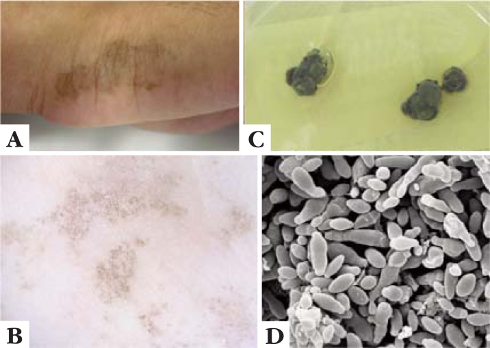
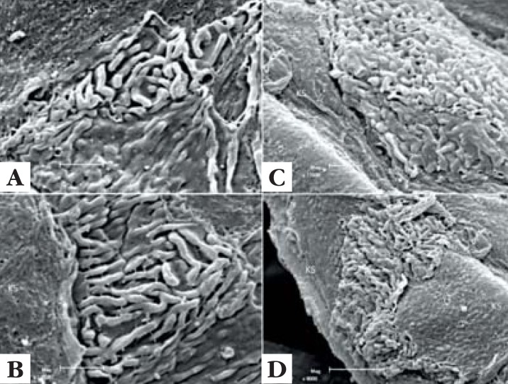
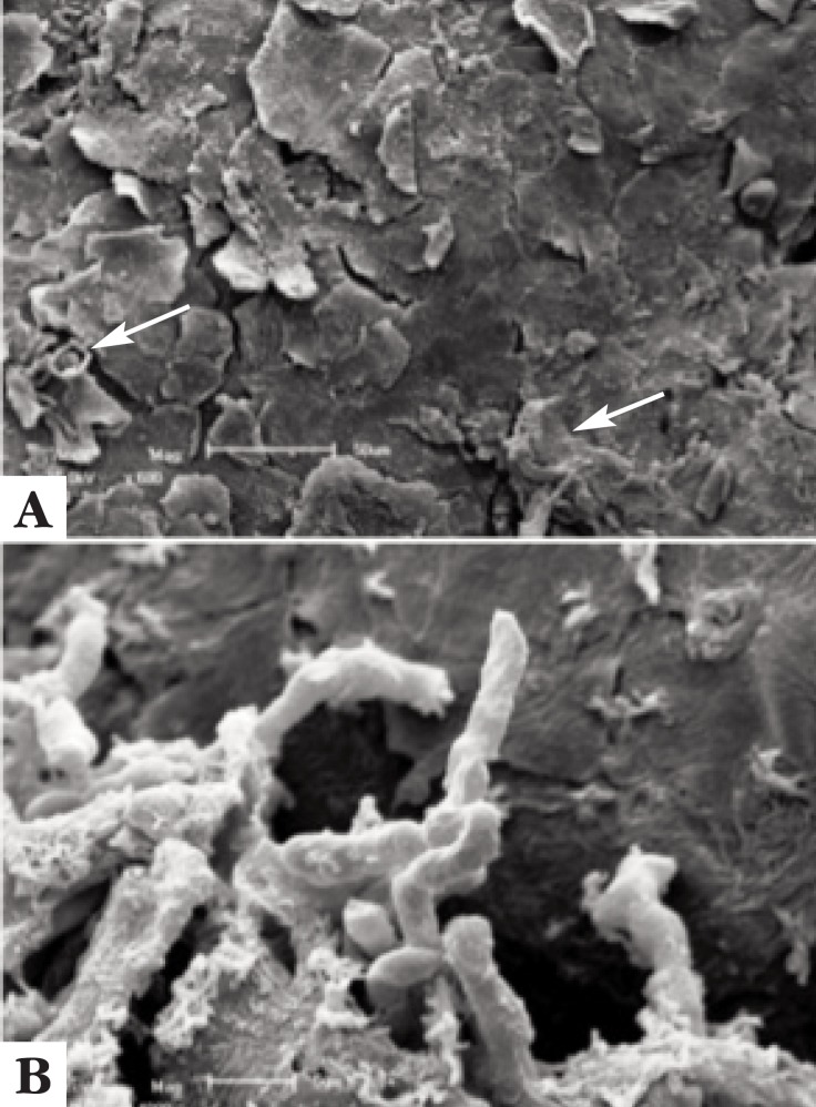
Tinea nigra is a rare superficial mycosis caused by Hortaea werneckii. This infection presents as asymptomatic brown to black maculae mostly in palmo-plantar regions. We performed scanning electron microscopy of a superficial shaving of a tinea nigra lesion. The examination of the outer surface of the sample showed the epidermis with corneocytes and hyphae and elimination of fungal filaments. The inner surface of the sample showed important aggregation of hyphae among keratinocytes, which formed small fungal colonies. The ultrastructural findings correlated with those of dermoscopic examination - the small fungal aggregations may be the dark spicules seen on dermoscopy - and also allowed to document the mode of dissemination of tinea nigra, showing how hyphae are eliminated on the surface of the lesion.
黑癣是一种由威尼克外瓶霉引起的罕见浅表真菌病。这种感染表现为大多在掌跖部位出现的无症状棕色至黑色斑疹。我们对黑癣病变的浅表刮屑进行了扫描电子显微镜检查。对样本外表面的检查显示表皮有角质形成细胞和菌丝,以及真菌丝的脱落。样本内表面显示角质形成细胞间有大量菌丝聚集,形成了小的真菌菌落。超微结构发现与皮肤镜检查结果相关——小的真菌聚集可能是皮肤镜下所见的深色刺——并且还能够记录黑癣的传播方式,显示菌丝在病变表面是如何脱落的。