Zhou Jiang-jun, MM, Department of Orthopedic, The 184th Hospital of Chinese PLA, Yingtan 335000, Jiangxi Province, China.
Zhao Min, MM, Department of Orthopedic, The 184th Hospital of Chinese PLA, Yingtan 335000, Jiangxi Province, China.
Pak J Med Sci. 2014 Mar;30(2):343-7.
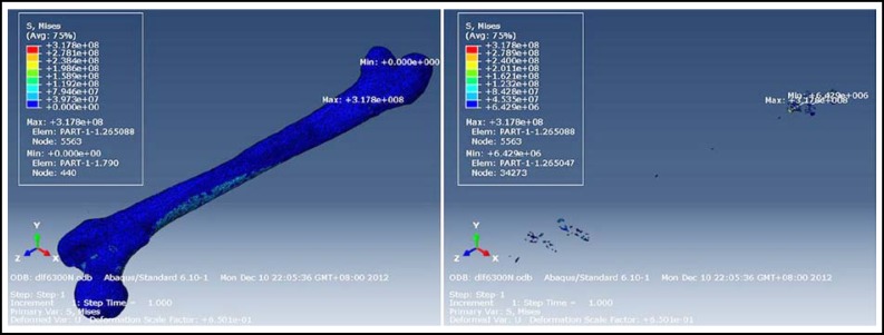
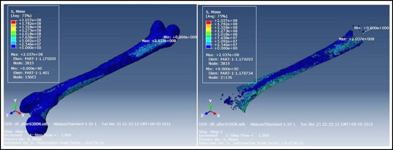
Finite element analysis was used to compare preoperative and postoperative stress distribution of a bone healing model of femur fracture, to identify whether broken ends of fractured bone would break or not after fixation dislodgement one year after intramedullary nailing. Method s: Using fast, personalized imaging, bone healing models of femur fracture were constructed based on data from multi-slice spiral computed tomography using Mimics, Geomagic Studio, and Abaqus software packages. The intramedullary pin was removed by Boolean operations before fixation was dislodged. Loads were applied on each model to simulate a person standing on one leg. The von Mises stress distribution, maximum stress, and its location was observed. Results : According to 10 kinds of display groups based on material assignment, the nodes of maximum and minimum von Mises stress were the same before and after dislodgement, and all nodes of maximum von Mises stress were outside the fracture line. The maximum von Mises stress node was situated at the bottom quarter of the femur. The von Mises stress distribution was identical before and after surgery. Conclusion : Fast, personalized model establishment can simulate fixation dislodgement before operation, and personalized finite element analysis was performed to successfully predict whether nail dislodgement would disrupt femur fracture or not.
使用有限元分析比较了股骨骨折骨愈合模型的术前和术后的应力分布,以确定髓内钉固定一年后骨折断端是否会在固定松动后断裂。方法:使用快速个性化成像技术,根据多层螺旋 CT 数据,使用 Mimics、Geomagic Studio 和 Abaqus 软件包构建股骨骨折骨愈合模型。在固定松动之前,通过布尔运算移除髓内钉。对每个模型施加负载以模拟一个人单腿站立。观察 von Mises 应力分布、最大应力及其位置。结果:根据基于材料分配的 10 种显示组,固定松动前后最大和最小 von Mises 应力的节点相同,并且所有最大 von Mises 应力的节点都在骨折线之外。最大 von Mises 应力节点位于股骨的底部四分之一处。手术前后 von Mises 应力分布相同。结论:快速个性化模型的建立可以在术前模拟固定松动,并且个性化有限元分析成功预测了钉松动是否会破坏股骨骨折。