Du Tao, Ju Guanqun, Wu Shuai, Cheng Zhongliang, Cheng Jun, Zou Xiangyu, Zhang Guangyuan, Miao Shuai, Liu Guohua, Zhu Yingjian
Department of Urology, Henan Provincial People's Hospital, Zhengzhou, P.R. China.
Department of Urology, Shanghai First People's Hospital, School of Medicine, Shanghai Jiao Tong University, Shanghai, P.R. China.
PLoS One. 2014 May 5;9(5):e96836. doi: 10.1371/journal.pone.0096836. eCollection 2014.
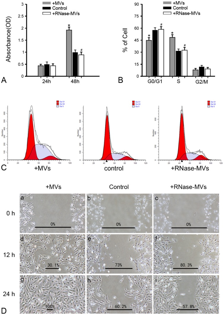
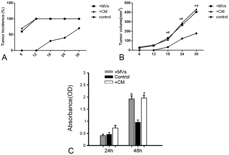
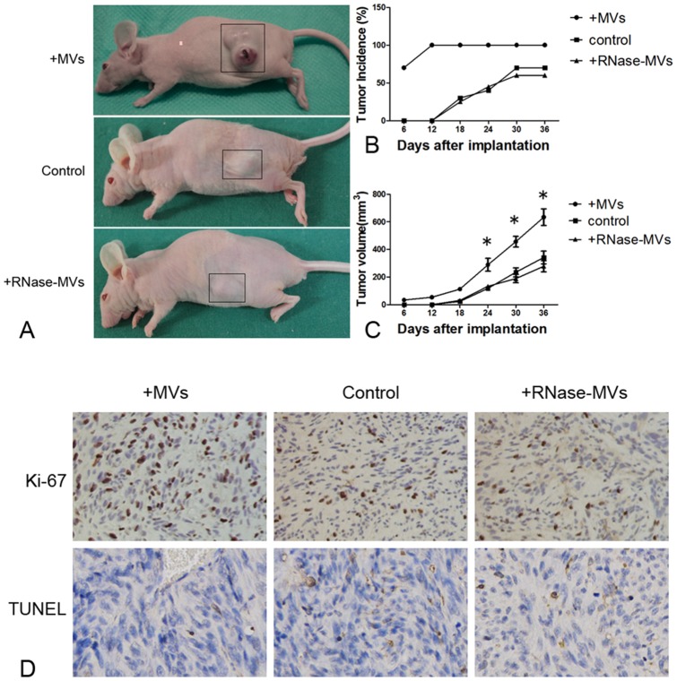
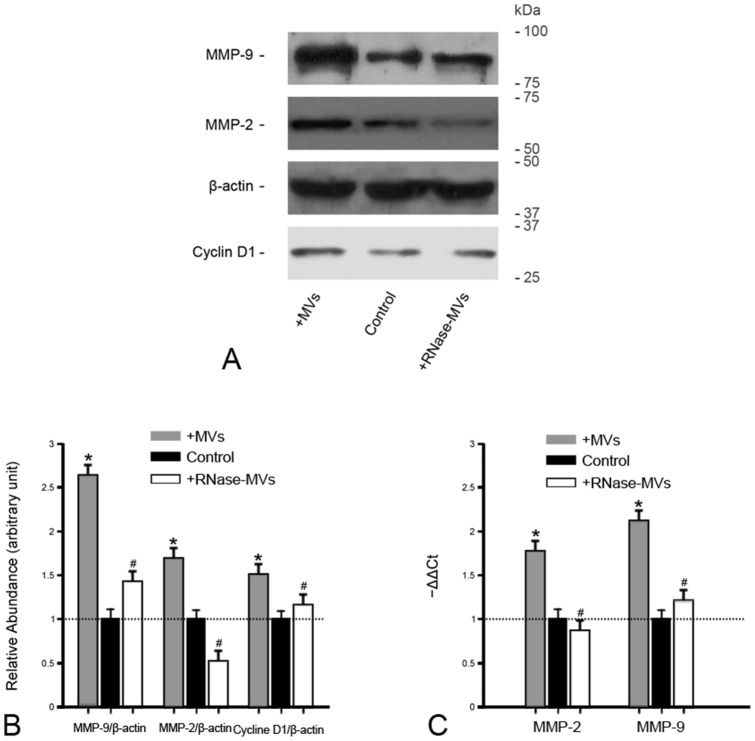
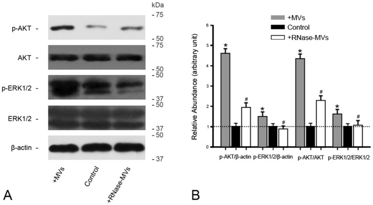
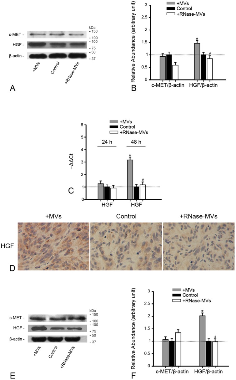
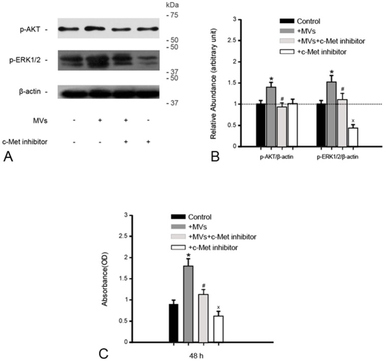
In our previous study, microvesicles (MVs) released from human Wharton's jelly mesenchymal stem cells (hWJ-MSCs) retard the growth of bladder cancer cells. We would like to know if MVs have a similar effect on human renal cell carcinoma (RCC). By use of cell culture and the BALB/c nu/nu mice xeno-graft model, the influence of MVs upon the growth and aggressiveness of RCC (786-0) was assessed. Cell counting kit-8 (CCK-8) assay, incidence of tumor, tumor size, Ki-67 or TUNEL staining was used to evaluate tumor cell growth in vitro or in vivo. Flow cytometry assay (in vitro) or examination of cyclin D1 expression (in vivo) was carried out to determine the alteration of cell cycle. The aggressiveness was analyzed by Wound Healing Assay (in vitro) or MMP-2 and MMP-9 expression (in vivo). AKT/p-AKT, ERK1/2/p-ERK1/2 or HGF/c-MET expression was detected by real-time PCR or western blot. Our data demonstrated that MVs promote the growth and aggressiveness of RCC both in vitro and in vivo. In addition, MVs facilitated the progression of cell cycle from G0/1 to S. HGF expression in RCC was greatly induced by MVs, associated with activation of AKT and ERK1/2 signaling pathways. RNase pre-treatment abrogated all effects of MVs. In summary, induction of HGF synthesis via RNA transferred by MVs activating AKT and ERK1/2 signaling is one of crucial contributors to the pro-tumor effect.
在我们之前的研究中,人脐带华通氏胶间充质干细胞(hWJ-MSCs)释放的微泡(MVs)可抑制膀胱癌细胞的生长。我们想了解MVs对人肾细胞癌(RCC)是否有类似作用。通过细胞培养和BALB/c裸鼠异种移植模型,评估了MVs对RCC(786-0)生长和侵袭性的影响。采用细胞计数试剂盒-8(CCK-8)检测、肿瘤发生率、肿瘤大小、Ki-67或TUNEL染色来评估体外或体内肿瘤细胞的生长情况。通过流式细胞术检测(体外)或细胞周期蛋白D1表达检测(体内)来确定细胞周期的改变。通过伤口愈合试验(体外)或MMP-2和MMP-9表达(体内)分析侵袭性。通过实时PCR或蛋白质印迹法检测AKT/p-AKT、ERK1/2/p-ERK1/2或HGF/c-MET的表达。我们的数据表明,MVs在体外和体内均促进RCC的生长和侵袭性。此外,MVs促进细胞周期从G0/1期向S期进展。MVs可显著诱导RCC中HGF的表达,这与AKT和ERK1/2信号通路的激活有关。核糖核酸酶预处理消除了MVs的所有作用。总之,MVs通过转移RNA诱导HGF合成,激活AKT和ERK1/2信号通路,是其促肿瘤作用的关键因素之一。