Varghese Frency, Bukhari Amirali B, Malhotra Renu, De Abhijit
Molecular Functional Imaging Laboratory, ACTREC, Tata Memorial Centre, Kharghar, Navi Mumbai, India.
PLoS One. 2014 May 6;9(5):e96801. doi: 10.1371/journal.pone.0096801. eCollection 2014.
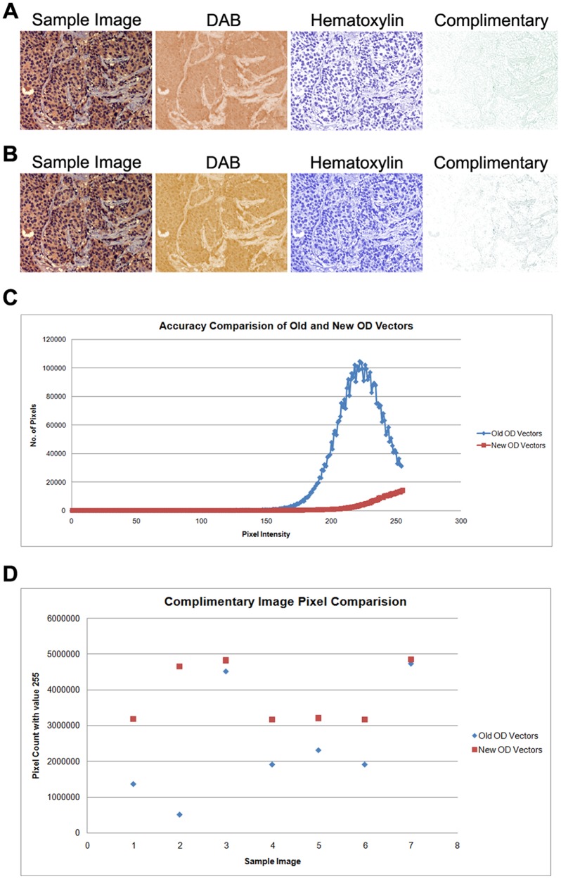
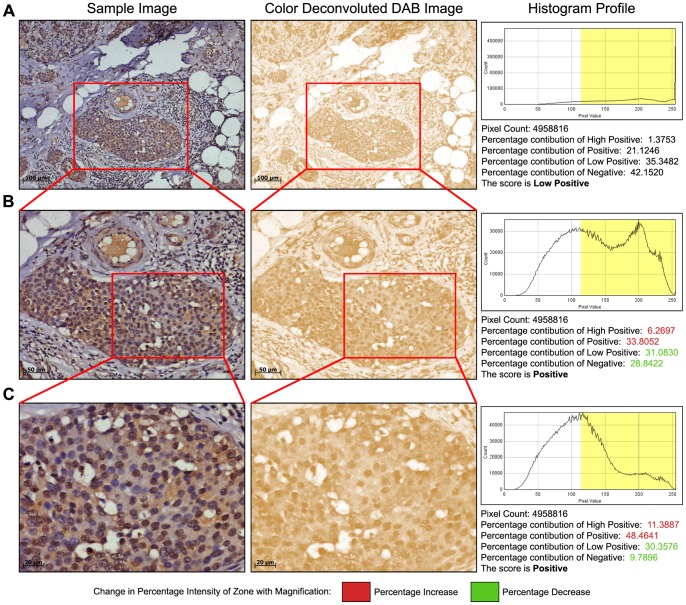
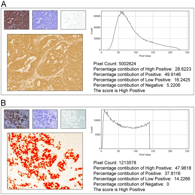
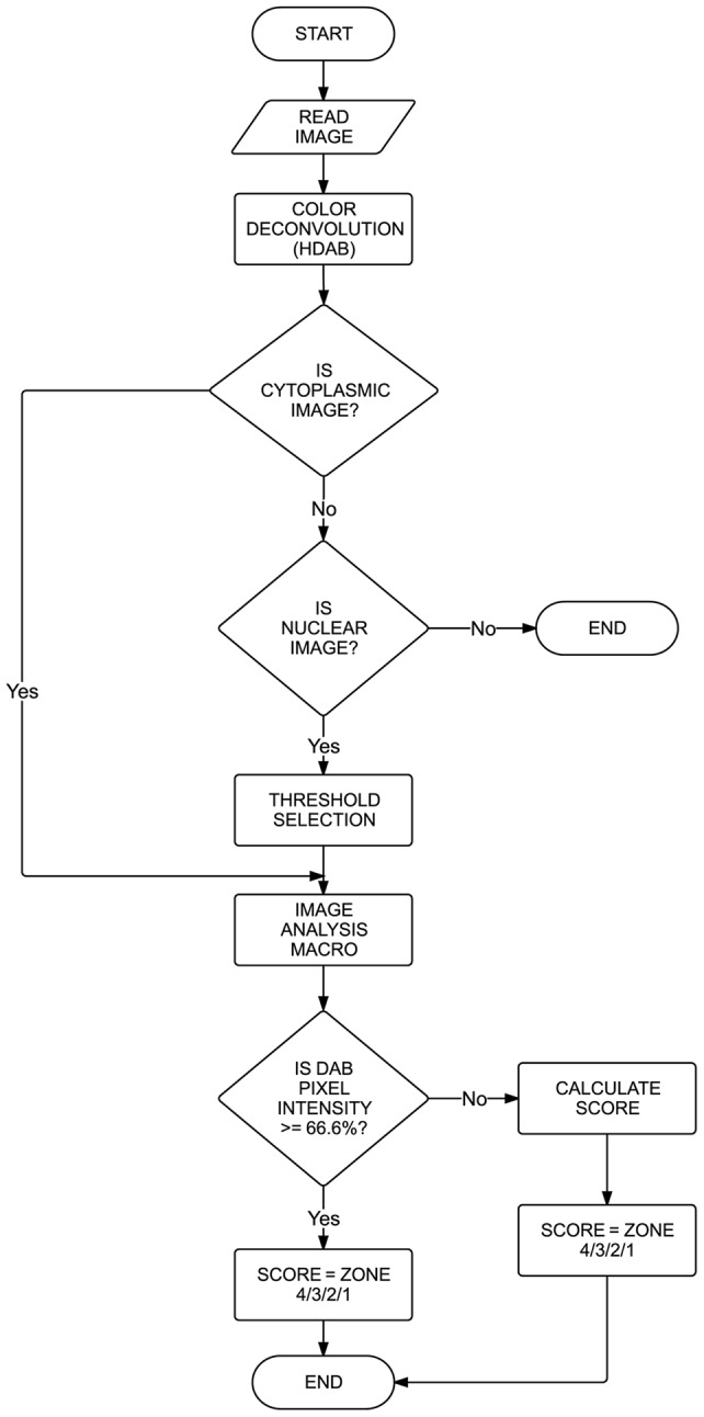
In anatomic pathology, immunohistochemistry (IHC) serves as a diagnostic and prognostic method for identification of disease markers in tissue samples that directly influences classification and grading the disease, influencing patient management. However, till today over most of the world, pathological analysis of tissue samples remained a time-consuming and subjective procedure, wherein the intensity of antibody staining is manually judged and thus scoring decision is directly influenced by visual bias. This instigated us to design a simple method of automated digital IHC image analysis algorithm for an unbiased, quantitative assessment of antibody staining intensity in tissue sections. As a first step, we adopted the spectral deconvolution method of DAB/hematoxylin color spectra by using optimized optical density vectors of the color deconvolution plugin for proper separation of the DAB color spectra. Then the DAB stained image is displayed in a new window wherein it undergoes pixel-by-pixel analysis, and displays the full profile along with its scoring decision. Based on the mathematical formula conceptualized, the algorithm is thoroughly tested by analyzing scores assigned to thousands (n = 1703) of DAB stained IHC images including sample images taken from human protein atlas web resource. The IHC Profiler plugin developed is compatible with the open resource digital image analysis software, ImageJ, which creates a pixel-by-pixel analysis profile of a digital IHC image and further assigns a score in a four tier system. A comparison study between manual pathological analysis and IHC Profiler resolved in a match of 88.6% (P<0.0001, CI = 95%). This new tool developed for clinical histopathological sample analysis can be adopted globally for scoring most protein targets where the marker protein expression is of cytoplasmic and/or nuclear type. We foresee that this method will minimize the problem of inter-observer variations across labs and further help in worldwide patient stratification potentially benefitting various multinational clinical trial initiatives.
在解剖病理学中,免疫组织化学(IHC)是一种用于识别组织样本中疾病标志物的诊断和预后方法,它直接影响疾病的分类和分级,进而影响患者的治疗管理。然而,直到如今在世界上大部分地区,组织样本的病理分析仍然是一个耗时且主观的过程,其中抗体染色强度是通过人工判断的,因此评分决策直接受到视觉偏差的影响。这促使我们设计一种简单的自动数字免疫组化图像分析算法,用于对组织切片中的抗体染色强度进行无偏倚的定量评估。第一步,我们通过使用颜色反卷积插件的优化光密度向量,采用DAB/苏木精颜色光谱的光谱反卷积方法,以正确分离DAB颜色光谱。然后,DAB染色的图像在一个新窗口中显示,在该窗口中对其进行逐像素分析,并显示完整的分析结果及其评分决策。基于所构思的数学公式,通过分析分配给数千张(n = 1703)DAB染色的免疫组化图像(包括从人类蛋白质图谱网络资源获取的样本图像)的分数,对该算法进行了全面测试。所开发的免疫组化分析器插件与开源数字图像分析软件ImageJ兼容,该软件创建数字免疫组化图像的逐像素分析概况,并进一步在四级系统中分配分数。手动病理分析与免疫组化分析器之间的比较研究结果显示匹配率为88.6%(P<0.0001,CI = 95%)。这种为临床组织病理学样本分析开发的新工具可在全球范围内用于对大多数标记蛋白表达为细胞质和/或核型的蛋白质靶点进行评分。我们预计,这种方法将最大限度地减少不同实验室之间观察者间差异的问题,并进一步有助于全球范围内的患者分层,可能使各种跨国临床试验计划受益。